Lung functions. Abstract: Lungs, their structure, topography and functions
Lungs, pulmones(from the Greek - pneumon, hence pneumonia - pneumonia), located in chest cavity, cavitas thoracis, on the sides of the heart and large vessels, in the pleural sacs, separated from each other mediastinum, mediastinum, extending from the spinal column behind to the front chest wall front.
Right lung larger volume than the left one (by approximately 10%), at the same time it is somewhat shorter and wider, firstly, due to the fact that the right dome of the diaphragm is higher than the left one (the influence of the voluminous right lobe liver), and secondly, the heart is located more to the left than to the right, thereby reducing the width of the left lung.
Every lung, pulmo, has an irregularly conical shape, with basis, basis pulmonis, directed downward, and a rounded apex, apex pulmonis, which stands 3 - 4 cm above the 1st rib or 2 - 3 cm above the clavicle in front, reaching level VII in the back cervical vertebra. At the top of the lungs a small groove is noticeable, sulcus subclavius, from the pressure passing here subclavian artery.
There are three surfaces in the lung. The lower one, fades diaphragmatica, is concave according to the convexity of the upper surface of the diaphragm to which it is adjacent. The extensive costal surface, fades costalis, is convex according to the concavity of the ribs, which, together with the intercostal muscles lying between them, form part of the wall of the thoracic cavity.
Medial surface, facies medialis, concave, repeats for the most part the outlines of the pericardium and is divided into the anterior part, adjacent to the mediastinum, pars mediastinal, and the posterior part, adjacent to the spinal column, pars vertebrdlis. The surfaces are separated by edges: the sharp edge of the base is called lower, margo inferior; the edge, also sharp, separating the fades medialis and costalis from each other, is margo anterior.
On medial surface located superiorly and posteriorly to the recess from the pericardium hilus pulmonis, through which the bronchi and pulmonary artery (as well as nerves) enter the lung, and two pulmonary veins (and lymphatic vessels) come out, putting it all together root easy-g oh, radix pulmonis. At the root lung bronchus located dorsally, the position of the pulmonary artery is different on the right and left sides. At the root of the right lung a. pulmonalis located below the bronchus, on the left side it crosses the bronchus and lies above it.
The pulmonary veins on both sides are located in the root of the lung below the pulmonary artery and bronchus. At the back, at the junction of the costal and medial surfaces of the lung, no sharp edge is formed; the rounded part of each lung is placed here in the recess of the chest cavity on the sides of the spine (sulci pulmonales).
Each lung through fissures, fissurae interlobares, divided by lobes, lobi. One furrow oblique, fissura obllqua, which is present on both lungs, begins relatively high (6-7 cm below the apex) and then obliquely descends down to the diaphragmatic surface, going deep into the substance of the lung.
She separates on every upper lung share of the bottom. In addition to this groove, the right lung also has a second, horizontal groove, fissura horizontalis, passing at the level of the IV rib. She delimits from upper lobe of the right lung is a wedge-shaped area that makes up the middle lobe. Thus, in the right lung there is three lobes: lobi superior, medius et inferior.
In the left lung, only two lobes are distinguished: superior, lobus superior, to which it goes apex of the lung, And lower, lobus inferior, more voluminous than the top one. It includes almost the entire diaphragmatic surface and most of the posterior obtuse margin of the lung. On the anterior edge of the left lung, in its lower part, there is cardiac notch, incisura cardiaca pulmonis sinistri, where the lung, as if pushed aside by the heart, leaves a significant part of the pericardium uncovered.
One of the most vital human organs is the lungs, which carry out the breathing process and ensure the exchange of oxygen and carbon dioxide in the body. In addition, they are involved in several other important processes in the body’s functioning and have a unique structure. In order to have a clear understanding of the work of this organ, you need to have a good knowledge of the anatomy of the lungs and their location. This paired organ consists of the left and right lungs.
Noticeably different from others internal organs, lung tissue has its own characteristic structure buildings. The name of this organ is due to its ability to remain on the surface of the water, due to the high volume of air in it. Latin name"pulmones" and the Greek "pneumon" also mean "lung". This is where the words “” (which heals) come from pulmonary diseases) and “pneumonia” (pulmonary inflammatory process).
The lungs are located in the chest cavity, occupying its main part (90%). The location and structure of the lungs makes it possible to unite all the important (main) vessels.
 Occupying almost the entire chest cavity, the lungs are located below with their base on the dome of the diaphragm. The right lower pulmonary section is separated by the diaphragm from the liver, the left – from the stomach, spleen, and part of the intestine. The median region is closely adjacent to the heart on both sides. The upper base is 4–5 cm above the collarbone.
The lungs are covered on the outside with serous tissue containment– pleura. On one side it passes into the pulmonary tissue, and on the other into the mediastinum and chest cavity. The resulting pleural cavity is filled with fluid. Due to this and due to the effect of negative pressure inside the cavity, the lung tissue is in a straightened state. The pleura located on the surface also protects the lungs from friction against the ribs during breathing.
The lungs are shaped like a cone, divided vertically into two parts. In this case, a convex surface and two concave ones are clearly visible. The convex pulmonary area (costal) is so close to the ribs that sometimes even the lung tissue has traces of them on the surface. One concave surface is located in the middle part of the body, and the second borders the diaphragm. In turn, each of them is also divided into interlobar sections.
 By appearance healthy lung tissue looks like a pink, finely porous sponge. Under the influence of unfavorable factors, its color changes - it becomes darker when age-related changes, pathologies, bad habits(smoking).
According to the anatomical structure, the lungs have different sizes, the right one is about 10% larger than the left one and they also differ in shape. The left one is smaller due to its “neighborhood” with the heart, which is closer to it, as if slightly displacing this area, called the cardiac notch. In this place, part of the pericardium remains unclosed, and there is a protrusion below, called the “pulmonary uvula”. The right lung is slightly higher than the left, due to the fact that the liver underneath it pushes it up a little.
On the medial side of each of them there is a “gate”. Through them important metabolic processes: the pulmonary artery, bronchi, and nerve plexuses enter the lungs, and the pulmonary veins and lymphatic vessels exit. Together, all this makes up the root of the lung. On the right, the pulmonary root is located behind the atrium and the superior vena cava, below the azygos vein, on the left - under the aortic arch.
Components of the lungs
The structure of the lungs is a complex structure, which consists of:
- bronchi;
- bronchioles;
- acini.

An important part of the respiratory system is the bronchi. These are tubular branches of the trachea that connect it to the lungs. Their main task– air passage. In shape they resemble the crown of a tree, due to the many branches and are called the “bronchial tree”. The bifurcation of the trachea into the left and right bronchi occurs in the area of the fifth thoracic vertebra. Then they enter the lung tissue and branch into lobar, then into segmental, and finally into the smallest channels - bronchioles.
Each pulmonary bronchus with the largest diameter has three membranes: 
- external;
- fibrinous-muscular, having cartilaginous tissue;
- located inside them mucus layer with ciliated epithelium.
With a decrease in the diameters of the branches of the bronchi, they cartilage tissue and mucous membrane gradually decrease. They are no longer present in the bronchioles, but cuboidal epithelium (a thin layer) is formed.
The framework of the lungs is the bronchial system, which has a branched structure. Many lobules measuring 15x25 mm make up each lung. The apices of the lobules include bronchioles (branches of the bronchi), at the ends of which there are acini - special formations covered a large number alveoli
Acini got their name because of their appearance, reminiscent of a bunch of grapes. Translated from Latin, Acinus means “bunch”. This is the main structural unit of lung tissue, which includes bronchioles, alveolar ducts, and primary pulmonary lobules in the form of small sacs.

The most important pulmonary elements are the alveoli, which ensure normal exchange of oxygen and carbon dioxide in the body. They are small thin-walled bubbles, tightly enveloped in a network of capillaries. Through the alveolar ducts, blood vessels are continuously supplied with oxygen and cleared of carbon dioxide. The tissue of each lung has 300 million alveoli. Oxygen is supplied to them by arterial capillaries, and carbon dioxide taken up by venous vessels.
The alveoli themselves are microscopic in size - 0.3 mm. But, thanks to their huge number, the average respiratory surface area when exhaling is 35 square meters, and when inhaling it can reach up to 100 square meters. Of course, the indicators depend on the person’s constitution - height, weight, fitness. Athletes have the highest marks.

From small structures Acini form lobules, then larger ones, of which the largest pulmonary area is formed - the lobe. The structure of the left and right lungs is different.
The right lung consists of three lobes:
- the upper of the three segments;
- the middle of the two segments;
- lower lobe of five segments.
The left lung consists of two lobes:
- top of five segments;
- the lower of the five segments.

Division into lobes occurs through grooves. One of them (oblique) begins at each lung below 6–7 cm from their apices and extends to the diaphragm, separating the upper lobe from the lower. In the right lung, in the region of the IV rib, there is a horizontal groove separating the wedge-shaped pulmonary area - the middle lobe.
The bronchopulmonary segments do not have clearly defined divisions. The pulmonary segment is a separate area into which blood flows from one artery and ventilation is provided by one bronchus (third order). Lung tissue is divided into segments different shapes, which differ not only in the right and left lungs, but are also located individually in each person.
Basic functions
In addition to the main respiratory function - ensuring gas exchange in the body, the lungs perform several other important missions:
- They normalize the pH composition in the blood, taking part in water, lipid, salt metabolism, and regulating the chlorine balance.
- Protect the body from respiratory infections, because they produce antimicrobial substances, immunoglobulins.
- Provide thermoregulation.
- Helps maintain normal water balance in the body.
- Participate in the creation of vocal sounds.
- They serve as a kind of blood storage (contain approximately 9% of the total volume).
- Protect the heart from mechanical influences.
- Promote the removal of toxins, essential and other compounds.
- Participate in coagulation (blood clotting).

The lungs are a paired human respiratory organ. The lungs are located in the chest cavity, adjacent to the right and left of the heart. They have the shape of a semi-cone, the base of which is located on the diaphragm, and the apex protrudes 1-3 cm above the collarbone. For prevention, drink Transfer Factor. The lungs are located in the pleural sacs, separated from each other by the mediastinum - a complex of organs that includes the heart, aorta, upper vena cava, extending from the spinal column at the back to the anterior chest wall at the front. They occupy most of the chest cavity and are in contact with both the spine and the anterior chest wall.
The right and left lungs are not the same in both shape and volume. The right lung has a larger volume than the left (by approximately 10%), at the same time it is somewhat shorter and wider due to the fact that the right dome of the diaphragm is higher than the left (the influence of the voluminous right lobe of the liver), and the heart is located more to the left, than to the right, thereby reducing the width of the left lung. In addition, on the right, directly under the lung in abdominal cavity there is a liver, which also reduces space.
The right and left lungs are located, respectively, in the right and left pleural cavities, or, as they are also called, pleural sacs. The pleura is a thin film consisting of connective tissue that covers the chest cavity from the inside (parietal pleura), and the lungs and mediastinum from the outside (visceral pleura). Between these two types of pleura there is a special lubricant that significantly reduces the friction force during breathing movements.
Each lung has an irregular conical shape with a base directed downward, its apex is rounded, it is located 3-4 cm above the first rib or 2-3 cm above the clavicle in front, and in the back it reaches the level of the VII cervical vertebra. At the top of the lungs, a small groove is noticeable, resulting from the pressure of the subclavian artery passing here. The lower border of the lungs is determined by percussion - tapping.
Both lungs have three surfaces: costal, inferior and medial (internal). Bottom surface has a concavity corresponding to the convexity of the diaphragm, and the costal ones, on the contrary, have a convexity corresponding to the concavity of the ribs from the inside. The medial surface is concave and basically follows the contours of the pericardium; it is divided into an anterior part adjacent to the mediastinum and a posterior part adjacent to the spinal column. The medial surface is considered the most interesting. Here, each lung has a so-called gate, through which the bronchus, pulmonary artery and vein enter the lung tissue.
The right lung consists of 3, and the left lung of 2 lobes. The skeleton of the lung is formed by tree-like branching bronchi. The boundaries of the lobes are deep grooves and are clearly visible. On both lungs there is an oblique groove, which begins almost at the apex, it is 6-7 cm below it, and ends at the lower edge of the lung. The groove is quite deep and represents the boundary between the upper and lower lobes of the lung. On the right lung there is an additional transverse groove that separates the middle lobe from the upper lobe. It is presented in the form of a large wedge. On the anterior edge of the left lung, in its lower part, there is a cardiac notch, where the lung, as if pushed aside by the heart, leaves a significant part of the pericardium uncovered. From below, this notch is limited by a protrusion of the anterior edge, called the uvula, adjacent to it part of the lung correspond middle share right lung.
In internal structure The lungs have a certain hierarchy that corresponds to the division of the main and lobar bronchi. According to the division of the lungs into lobes, each of the two main bronchi, approaching the gates of the lung, begins to divide into lobar bronchi. The right upper lobar bronchus, heading towards the center of the upper lobe, passes over the pulmonary artery and is called supraarterial, the remaining lobar bronchi of the right lung and all the lobar bronchi of the left pass under the artery and are called subarterial. The lobar bronchi, penetrating into the substance of the lung, are divided into smaller tertiary bronchi, called segmental, since they ventilate specific areas of the lung - segments. Each lobe of the lung consists of several segments. The segmental bronchi, in turn, are divided dichotomously (each into two) into smaller bronchi of the 4th and subsequent orders up to the terminal and respiratory bronchioles.
Each lobe or segment receives blood supply from its own branch of the pulmonary artery, and the outflow of blood is also carried out through a separate inflow pulmonary vein. Vessels and bronchi always pass through the thickness of the connective tissue, which is located between the lobules. Secondary lobules of the lung - so named to distinguish them from the primary lobules, which are smaller. Correspond to the branches of the lobar bronchi.
The primary lobule is the entire set of pulmonary alveoli, which is associated with the smallest bronchiole of the last order. The alveolus is the final section of the respiratory tract. In fact, the lung tissue itself consists of alveoli. They look like tiny bubbles, and neighboring ones have common walls. The inside walls of the alveoli are covered epithelial cells, which are of two types: respiratory (respiratory alveocytes) and large alveocytes. Respiratory cells very highly specialized cells that perform the function of gas exchange between the environment and the blood. Large alveocytes produce a specific substance - surfactant. Lung tissue always contains a certain number of phagocytes - cells that destroy foreign particles and small bacteria.
The main function of the lungs is gas exchange, when the blood is enriched with oxygen and carbon dioxide is removed from the blood. The entry of oxygen-saturated air into the lungs and the removal of exhaled, carbon dioxide-saturated air to the outside are ensured by active respiratory movements of the chest wall and diaphragm and contractility the easiest in combination with the activity of the respiratory tract. Unlike other parts of the respiratory tract, the lungs do not transport air, but directly carry out the transition of oxygen into the blood. This occurs through the membranes of the alveoli and respiratory alveocytes. In addition to normal breathing in the lung, there is collateral breathing, i.e., the movement of air bypassing the bronchi and bronchioles. It occurs between the peculiarly constructed acini, through pores in the walls of the pulmonary alveoli.
The physiological role of the lungs is not limited to gas exchange. Their complex anatomical structure also corresponds to a variety of functional manifestations: activity of the bronchial wall during breathing, secretory-excretory function, participation in metabolism (water, lipid and salt with the regulation of chlorine balance), which is important in maintaining acid-base balance in the body.
It is interesting to note that the blood supply to the lungs is dual, as they have two completely independent vascular networks. One of them is responsible for breathing and comes from the pulmonary artery, and the second provides the organ with oxygen and comes from the aorta. Venous blood flowing to the pulmonary capillaries through the branches of the pulmonary artery enters into osmotic exchange (gas exchange) with the air contained in the alveoli: it releases its carbon dioxide into the alveoli and receives oxygen in return. Arterial blood brought to the lungs from the aorta. It nourishes the wall of the bronchi and lung tissue.
In the lungs there are superficial lymphatic vessels embedded in deep layer pleura, and deep, inside pulmonary. The roots of the deep lymphatic vessels are the lymphatic capillaries, which form networks around the respiratory and terminal bronchioles, in the interacini and interlobular septa. These networks continue into the plexuses of lymphatic vessels around the branches of the pulmonary artery, veins and bronchi.
While a person is alive, he breathes. What is breathing? These are processes that continuously supply all organs and tissues with oxygen and remove carbon dioxide from the body, which is formed as a result of the metabolic system. Performs these vital processes which directly interact with the cardiovascular system. To understand how gas exchange occurs in the human body, you should study the structure and functions of the lungs.
Why does a person breathe?
The only way to obtain oxygen is through breathing. It is not possible to hold it for a long time, since the body requires another portion. Why do we need oxygen at all? Without it, metabolism will not occur, the brain and all other human organs will not work. With the participation of oxygen they split nutrients, energy is released, and every cell is enriched with them. Breathing is commonly called gas exchange. And rightly so. After all, the peculiarities of the respiratory system are to take in oxygen from the air that enters the body and remove carbon dioxide.
What are human lungs
Their anatomy is quite complex and variable. This organ is paired. Its location is the chest cavity. The lungs are adjacent to the heart on both sides - right and left. Nature has made sure that both of these most important organ were protected from compression, impacts, etc. In front, the barrier to damage is the back - the spinal column, and on the sides - the ribs.
The lungs are literally riddled with hundreds of branches of bronchi, with alveoli the size of a pinhead located at their ends. Them in the body healthy person there are up to 300 million pieces. The alveoli play an important role: they supply blood vessels with oxygen and, having a branched system, are able to provide for gas exchange large area. Just imagine: they can cover the entire surface of a tennis court!
In appearance, the lungs resemble semi-cones, the bases of which are adjacent to the diaphragm, and the tops with rounded ends protrude 2-3 cm above the collarbone. The human lungs are a rather unique organ. The anatomy of the right and left lobes is different. So, the first one is slightly larger in volume than the second one, while it is somewhat shorter and wider. Each half of the organ is covered with pleura, consisting of two layers: one is fused with the chest, the other with the surface of the lung. The outer pleura contains glandular cells, thanks to which pleural cavity fluid is produced.
The inner surface of each lung has a depression called the hilum. They include the bronchi, the base of which looks like a branching tree, and the pulmonary artery, and a pair of pulmonary veins emerge.
Human lungs. Their functions
Of course, there are no secondary organs in the human body. The lungs are also important in ensuring human life. What kind of work do they do?
- The main functions of the lungs are to carry out respiratory process. A person lives while he breathes. If the supply of oxygen to the body is cut off, death will occur.
- The work of the human lungs is to remove carbon dioxide, due to which the body maintains acid-base balance. Through these organs, a person gets rid of volatile substances: alcohol, ammonia, acetone, chloroform, ether.

- The functions of the human lungs do not end there. The paired organ is still involved in which comes into contact with air. As a result, an interesting thing happens chemical reaction. Oxygen molecules in the air and carbon dioxide molecules in dirty blood change places, i.e. oxygen replaces carbon dioxide.
- The various functions of the lungs allow them to participate in the water exchange occurring in the body. Up to 20% of the liquid is removed through them.
- The lungs are active participants in the process of thermoregulation. They release 10% of their heat into the atmosphere when they exhale.
- Regulation is not complete without the participation of the lungs in this process.
How do the lungs work?
The functions of the human lungs are to transport the oxygen contained in the air into the blood, use it, and remove carbon dioxide from the body. The lungs are fairly large soft organs with spongy tissue. The inhaled air enters the air sacs. They are separated from each other by thin walls with capillaries.
There are only small cells between the blood and the air. Therefore, thin walls do not create obstacles for inhaled gases, which facilitates good passage through them. In this case, the functions of the human lungs are to use necessary and remove unnecessary gases. Lung tissue is very elastic. When you inhale, expansion occurs chest and an increase in lung volume.
The windpipe, represented by the nose, pharynx, larynx, trachea, looks like a tube 10-15 cm long, divided into two parts called bronchi. Air passing through them enters the air sacs. And when you exhale, the volume of the lungs decreases, the chest decreases in size, and the pulmonary valve partially closes, which allows air to escape again. This is how human lungs work.

Their structure and functions are such that the capacity of this organ is measured by the amount of inhaled and exhaled air. So, for men it is equal to seven pints, for women - five. The lungs are never empty. The air remaining after exhalation is called residual air. When you inhale, it mixes with fresh air. Therefore, breathing is a conscious and at the same time unconscious process that occurs constantly. A person breathes when he sleeps, but he does not think about it. In this case, if you wish, you can interrupt your breathing for a short time. For example, while underwater.
Interesting facts about lung function
They are capable of pumping 10 thousand liters of inhaled air per day. But it is not always crystal clear. Along with oxygen, dust, many microbes and foreign particles enter our body. Therefore, the lungs perform the function of protection from all unwanted impurities in the air.
The walls of the bronchi have many tiny villi. They are needed to trap germs and dust. And the mucus, which is produced by the cells of the walls of the respiratory tract, lubricates these villi, and is then expelled when coughing.
It consists of organs and tissues that are to the fullest provide ventilation and breathing. The functions of the respiratory system lie in the implementation of gas exchange - the main link in metabolism. The latter is responsible only for pulmonary (external) respiration. It includes:
1. consisting of the nose and its cavity, larynx, trachea, bronchi.
The nose and its cavity heat, humidify and filter the inhaled air. Its cleansing is achieved through numerous hard hairs and goblet cells with cilia.
The larynx is located between the root of the tongue and the trachea. Its cavity is divided by the mucous membrane in the form of two folds. They are not completely fused in the middle. The gap between them is called the glottis.
The trachea originates from the larynx. In the chest it is divided into bronchi: right and left.
2. Lungs with densely branched vessels, bronchioles and alveolar sacs. They begin the gradual division of the main bronchi into small tubes called bronchioles. They make up the smallest structural lung elements- lobules.
The pulmonary artery carries blood from the right ventricle of the heart. It is divided into left and right. The branching of the arteries follows the bronchi, entwining the alveoli and forming small capillaries.
3. Musculoskeletal system, thanks to which a person is not limited in breathing movements.
These are the ribs, muscles, diaphragm. They monitor the integrity of the airways and maintain them during various postures and body movements. Muscles, contracting and relaxing, contribute to changes. The diaphragm is designed to separate the thoracic cavity from the abdominal cavity. It is the main muscle involved in normal inhalation.
A man breathes through his nose. Next, the air passes through the airways and enters the human lungs, the structure and functions of which ensure the further functioning of the respiratory system. This is a purely physiological factor. This type of breathing is called nasal breathing. In the cavity of this organ, heating, humidification and purification of the air occurs. If the nasal mucosa is irritated, the person sneezes and protective mucus begins to be released. Nasal breathing may be difficult. Then the air enters the throat through the mouth. Such breathing is said to be oral and, in fact, pathological. In this case, the functions of the nasal cavity are disrupted, which causes various diseases respiratory tract.

From the pharynx, air is directed to the larynx, which performs other functions besides carrying oxygen further into the respiratory tract, in particular, reflexogenic. If irritation occurs of this body, a cough or spasm appears. In addition, the larynx is involved in sound production. This is important for any person, since his communication with other people occurs through speech. They continue to heat and humidify the air, but this is not their main function. By performing certain work, they regulate the volume of inhaled air.
Respiratory system. Functions
The air around us contains oxygen, which can penetrate into our body through the skin. But its quantity is not enough to support life. This is why the respiratory system exists. Transportation of necessary substances and gases is carried out by circulatory system. The structure of the respiratory system is such that it is able to supply the body with oxygen and remove carbon dioxide from it. It performs the following functions:
- Regulates, conducts, humidifies and degreases the air, removes dust particles.
- Protects the respiratory tract from food particles.
- Carries air into the trachea from the larynx.
- Improves gas exchange between the lungs and blood.
- Transports venous blood to the lungs.
- Saturates blood with oxygen and removes carbon dioxide.
- Performs a protective function.
- Detains and resolves blood clots, particles of foreign origin, emboli.
- Performs the metabolism of necessary substances.
An interesting fact is that with age there is a limitation functionality respiratory system. The level of ventilation of the lungs and the work of breathing decreases. The causes of such disorders can be various changes in the bones and muscles of a person. As a result, the shape of the chest changes and its mobility decreases. This leads to a decrease in the capabilities of the respiratory system.
Breathing phases
When you inhale, oxygen from the alveoli of the lungs enters the blood, namely the red blood cells. From here, on the contrary, carbon dioxide passes into the air, which contained oxygen. From the moment air enters until air leaves the lungs, its pressure in the organ increases, which stimulates the diffusion of gases.
When you exhale, a pressure greater than atmospheric pressure is created in the alveoli of the lungs. The diffusion of gases: carbon dioxide and oxygen begins to take place more actively.
Every time after exhalation a pause is created. This happens because there is no diffusion of gases, since the pressure of the air remaining in the lungs is insignificant, much lower than atmospheric pressure.
As long as I breathe, I live. Breathing process
- The baby in the womb receives oxygen through her blood, so the baby's lungs do not take part in the process; they are filled with fluid. When a baby is born and takes its first breath, the lungs begin to work. The structure and functions are such that they are able to provide the human body with oxygen and remove carbon dioxide.
- Signals about the amount of oxygen required at a specific period of time are given by respiratory center, which is located in the brain. Thus, during sleep, much less oxygen is required than during working hours.
- The volume of air entering the lungs is regulated by messages sent by the brain.

- When this signal arrives, the diaphragm expands, which leads to stretching of the chest. This maximizes the volume that the lungs occupy when they expand during inhalation.
- During exhalation, the diaphragm and intercostal muscles relax, and the volume of the chest decreases. This causes air to be pushed out of the lungs.
Types of breathing
- Clavicular. When a person hunches, his shoulders are raised and his stomach is compressed. This indicates insufficient oxygen supply to the body.
- Chest breathing. It is characterized by expansion of the chest due to the intercostal muscles. Such functions help saturate the body with oxygen. This method, purely physiologically, is more suitable for pregnant women.
- Deep breathing fills the air lower sections organs. Most often, athletes and men breathe this way. This method is convenient during physical activity.
It is not without reason that they say that breathing is a mirror of mental health. Thus, the psychiatrist Lowen noticed an amazing relationship between character and type emotional disorder person. In people prone to schizophrenia, breathing involves the upper chest. And a person with a neurotic type of character breathes more with his stomach. Usually people use mixed breathing, which involves both the chest and the diaphragm.
Lungs of people who smoke
Smoking causes swipe by organs. Tobacco smoke contains tar, nicotine and hydrogen cyanide. These harmful substances have the ability to settle on the lung tissue, resulting in the death of the organ epithelium. The lungs of a healthy person are not subject to such processes.
People who smoke have lungs that are dirty gray or black in color due to congestion. huge amount dead cells. But these are not all negative aspects. Lung functions are significantly reduced. Negative processes begin, leading to inflammation. As a result, a person suffers from chronic obstructive pulmonary diseases, which contribute to the development respiratory failure. It, in turn, causes numerous disorders that occur due to a lack of oxygen in the tissues of the body.

Social advertising constantly shows clips and pictures with the difference between the lungs of a healthy person and a smoker. And many people who have never picked up a cigarette breathe a sigh of relief. But you shouldn’t get your hopes up too much, thinking that the terrible sight that is the lungs of a smoker has nothing to do with you. The interesting thing is that at first glance there is no particular external difference. Neither x-ray, nor ordinary fluorography will show whether the person being examined smokes or not. Moreover, not a single pathologist can determine with absolute certainty whether a person had an addiction to smoking during life until he detects typical signs: the condition of the bronchi, yellowing of the fingers, and so on. Why? It turns out that harmful substances floating in the polluted air of cities, entering our body, just like tobacco smoke, enter the lungs...
The structure and functions of this organ are designed to protect the body. It is known that toxins destroy lung tissue, which subsequently, due to the accumulation of dead cells, acquires a dark color.
Interesting things about breathing and the respiratory system
- The lungs are the size of a human palm.
- Volume paired organ- 5 liters. But it is not fully used. To ensure normal breathing 0.5 l is enough. The volume of residual air is one and a half liters. If you count, then exactly three liters of air volume are always in reserve.
- The older a person is, the less frequent his breathing. In one minute, a newborn inhales and exhales thirty-five times, a teenager twenty, an adult fifteen times.
- In one hour a person takes a thousand breaths, in a day - twenty-six thousand, in a year - nine million. Moreover, men and women do not breathe the same way. In one year, the former take 670 million inhalations and exhalations, and the latter - 746.
- In one minute, it is vital for a person to receive eight and a half liters of air volume.
Based on all of the above, we conclude: you need to take care of your lungs. If you have any doubts about the health of your respiratory system, consult your doctor.
The lungs (pulmones) represent the main respiratory organs, filling the entire chest cavity except the mediastinum. Gas exchange occurs in the lungs, that is, oxygen is absorbed from the air of the alveoli by red blood cells and carbon dioxide is released, which in the lumen of the alveoli breaks down into carbon dioxide and water. Thus, in the lungs there is a close connection of the airways, blood and lymphatic vessels and nerves. The combination of paths for carrying air and blood in a special respiratory system can be traced with early stages embryonic and phylogenetic development. The supply of oxygen to the body depends on the degree of ventilation of various parts of the lungs, the relationship between ventilation and the speed of blood flow, the saturation of blood with hemoglobin, the rate of diffusion of gases through the alveolar capillary membrane, the thickness and elasticity of the elastic frame of the lung tissue, etc. A change in at least one of these indicators leads to disruption of respiratory physiology and may cause certain functional impairments.
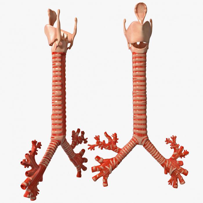
303. Larynx, trachea and lungs in front.
1 - larynx; 2 - trachea; 3 - apex pulmonis; 4 - facies costalis; 5 - lobus superior; 6 - pulmo sinister; 7 - fissura obliqua; 8 - lobus inferior; 9 - basis pulmonis; 10 - lingula pulmonis; 11 - impressio cardiaca; 12 - margo posterior; 13 - margo anterior; 14 - facies diaphragmatica; 15 - margo inferior; 16 - lobus inferior; 17 - lobus medius; 18 - fissura horizontalis; 19 - pulmo dexter; 20 - lobus superior; 21 - bifurcatio tracheae.
The external structure of the lungs is quite simple (Fig. 303). The shape of the lung resembles a cone, where there is an apex (apex), base (basis), costal convex surface (facies costalis), diaphragmatic surface (facies diaphragmatica) and medial surface (facies medialis). The last two surfaces are concave (Fig. 304). On the medial surface, the vertebral part (pars vertebralis), the mediastinal part (pars mediastinalis) and the cardiac impression (impressio cardiaca) are distinguished. The left deep cardiac depression is complemented by a cardiac notch (incisura cardiaca). In addition, there are interlobar surfaces (facies interlobares). The anterior edge (margo anterior), separating the costal and medial surfaces, is distinguished; the lower edge (margo inferior) is at the junction of the costal and diaphragmatic surfaces. The lungs are covered with a thin visceral layer of pleura, through which the darker areas of connective tissue located between the bases of the lobules are visible. On the medial surface, the visceral pleura does not cover the hilus pulmonum, but descends below them in the form of a duplication called the pulmonary ligaments (ligg. pulmonalia).

304. Mediastinal surface and root of the right lung. 1 - apex pulmonis; 2 - place of transition of the pleura from the visceral layer to the mediastinal layer; 3 - aa. pulmonales; 4 - bronchus principalis; 5 - vv. pulmonales; 6 - lig. pulmonary

305. Mediastinal surface and root of the left lung. 1 - apex pulmonis; 2 - place of transition of the pleura from the visceral layer to the mediastinal layer; 3 - aa. pulmonales; 4 - bronchus principalis; 5 - v. pulmonalis.
At the gate of the right lung, the bronchus is located above, then the pulmonary artery and vein (Fig. 304). In the left lung there is a pulmonary artery at the top, then a bronchus and a vein (Fig. 305). All these formations form the root of the lungs (radix pulmonum). The root of the lung and the pulmonary ligament hold the lungs in a certain position. On the costal surface of the right lung there is a horizontal fissure (fissura horizontalis) and below it an oblique fissure (fissura obliqua). The horizontal fissure is located between the linea axillaris media and the linea sternalis of the chest and coincides with the direction of the IV rib, and the oblique fissure with the direction of the VI rib. At the back, starting from the linea axillaris to the linea vertebralis of the chest, there is one groove, representing a continuation of the horizontal groove. Due to these grooves in the right lung, the upper, middle and lower lobes are distinguished (lobi superior, medius et inferior). The largest lobe is the lower one, then comes the upper and middle - the smallest. In the left lung there are upper and lower lobes, separated by a horizontal fissure. Below the cardiac notch on the anterior edge there is a tongue (lingula pulmonis). This lung is slightly longer than the right one, which is due to the lower position of the left dome of the diaphragm.
Boundaries of the lungs. The tops of the lungs protrude onto the neck above the collarbone by 3-4 cm.
The lower border of the lungs is determined at the point of intersection of the rib with conditionally drawn lines on the chest: along the linea parasternalis - VI rib, along the linea medioclavicularis (mamillaris) - VII rib, along the linea axillaris media - VIII rib, along the linea scapularis - X rib, along the linea paravertebralis - at the head of the XI rib.
With maximum inspiration, the lower edge of the lungs, especially along the last two lines, drops by 5 - 7 cm. Naturally, the border of the visceral layer of the pleura coincides with the border of the lungs.
The anterior edge of the right and left lungs is projected onto the anterior surface of the chest differently. Starting from the apexes of the lungs, the edges run almost parallel at a distance of 1 -1.5 cm from each other to the level of the cartilage of the 4th rib. In this place, the edge of the left lung deviates to the left by 4-5 cm, leaving the cartilage of the IV-V ribs not covered by the lung. This cardiac impression (impressio cardiaca) is filled with the heart. The anterior edge of the lungs at the sternal end of the VI rib passes into the lower edge, where the boundaries of both lungs coincide.
Internal structure of the lungs. Lung tissue divided into non-parenchymal and parenchymal components. The first includes all bronchial branches, branches of the pulmonary artery and pulmonary vein (except for capillaries), lymphatic vessels and nerves, connective tissue layers lying between the lobules, around the bronchi and blood vessels, as well as the entire visceral pleura. The parenchymal part consists of alveoli - alveolar sacs and alveolar ducts with surrounding blood capillaries.
306. Scheme of the order of generation of branching of the bronchi in the lung lobule.
1 - trachea; 2 - bronchus principalis; 3 - bronchus lobaris; 4 - bronchus segmentalis; 5, 6 - intermediate bronchi; 7 - bronchus interlobularis; 8 - bronchus terminalis; 9 - bronchioli I; 10 - bronchioli II; 11-13 bronchioli respiratorii I, II, III; 14 - alveoli with alveolar ducts, connected to form an acini; 15 - transitory zone; 16 - respiratory zone.
Bronchial architecture(Fig. 306). The right and left pulmonary bronchi at the hilum of the lungs are divided into lobar bronchi (bronchi lobares). All lobar bronchi pass under the large branches of the pulmonary artery, with the exception of the right upper lobe bronchus, which is located above the artery. The lobar bronchi are divided into segmental bronchi, which are successively divided in the form of an irregular dichotomy up to the 13th order, ending with a lobular bronchus (bronchus lobularis) with a diameter of about 1 mm. Each lung has up to 500 lobular bronchi. The wall of all bronchi contains cartilaginous rings and spiral plates, reinforced with collagen and elastic fibers and alternating with muscle elements. In the mucous membrane of the bronchial tree, mucous glands are richly developed (Fig. 307).

307. Transverse section of a segmental bronchus.
1 - cartilage; 2 - mucous glands; 3 - fibrous connective tissue with muscle elements; 4 - mucous membrane.
When the lobular bronchus divides, a qualitatively new formation arises - the terminal bronchi (bronchi terminales) with a diameter of 0.3 mm, which are already devoid of a cartilaginous base and are lined with a single layer prismatic epithelium. The terminal bronchi, sequentially dividing, form bronchioles of the 1st and 2nd order (bronchioli), in the walls of which there is a well-developed muscle layer, capable of blocking the lumen of bronchioles. They, in turn, are divided into respiratory bronchioles of the 1st, 2nd and 3rd order (bronchioli respiratorii). Respiratory bronchioles are characterized by the presence of communications directly with the alveolar ducts (Fig. 308). Respiratory bronchioles of the 3rd order communicate with 15-18 alveolar ducts (ductuli alveolares), the walls of which are formed by alveolar sacs (sacculi alveolares) containing alveoli (alveoli). The branching system of the 3rd order respiratory bronchiole develops into the lung acinus (Fig. 306).
The structure of the alveoli. As mentioned above, the alveoli are part of the parenchyma and represent the final part of the air system where gas exchange takes place. The alveoli represent a protrusion of the alveolar ducts and sacs (Fig. 308). They have a cone-shaped base with an elliptical cross-section (Fig. 309). There are up to 300 million alveoli; they make up a surface equal to 70-80 m2, but the respiratory surface, i.e., the places of contact between the capillary endothelium and the alveolar epithelium, is smaller and equal to 30-50 m2. Alveolar air is separated from the blood capillaries by a biological membrane that regulates the diffusion of gases from the cavity of the alveoli into the blood and back. The alveoli are covered with small, large and loose flat cells. The latter are also able to phagocytose foreign particles. These cells are located on the basement membrane. The alveoli are surrounded by blood capillaries, their endothelial cells are in contact with the alveolar epithelium. Gas exchange occurs at the sites of these contacts. The thickness of the endothelial-epithelial membrane is 3-4 microns.

308. Histological section of the lung parenchyma of a young woman showing multiple alveoli (A) that are partially connected to the alveolar duct (AD) or respiratory bronchiole (RB). RA - branch of the pulmonary artery, x 90 (according to Weibel).

309. Section of the lung (A). Two alveoli (1) are visible, open from the side of the alveolar duct (2). Schematic model of the location of the alveoli around the alveolar duct (B) (according to Weibel).
Between basement membrane capillary and the basement membrane of the alveolar epithelium, there is an interstitial zone containing elastic, collagen fibers and the finest fibrils, macrophages and fibroblasts. Fibrous formations give elasticity to the lung tissue; due to it, the act of exhalation is ensured.


