Olfactory histology. I
II. Organ of vision.
III. Organs of hearing and balance.
IV. Organs of smell and taste.
I. Functions and classification of sense organs
The body’s perception of information about the state of the external and internal environment, as well as its processing and transformation into sensations, is carried out by analyzers. They are complex structural and functional systems that connect the central nervous system with the external and internal environment. Each analyzer consists of three parts: a peripheral one, in which the perception of stimuli occurs, an intermediate one, represented by pathways and subcortical formations, and a central one, represented by areas of the cerebral cortex, where the analysis of information and the synthesis of sensations occurs.
The sense organs are the primary parts of the analyzers. They contain special receptor cells of two types: neurosensory and sensoroepithelial. Neurosensory cells develop from neuroectoderm. They are called primary senses. Sense organs with nerve receptor cells belong to group I (organs of vision and smell). Sensoepithelial receptor cells perceive irritation and transmit them to the dendrites of sensory neurons, which perceive excitation and form a nerve impulse. Sensoepithelial receptor cells originate from the dermal actoderm and are called secondary sensory cells. Sense organs with such receptor cells belong to group II (organs of hearing, balance, taste).
The peripheral parts of the third group of sensory organs include encapsulated and non-encapsulated nerve endings. All of them are dendrites of sensory ganglion neurons. Based on their function, exteroreceptors and interoreceptors are distinguished. Exteroreceptors perceive irritation from the external environment. These are tactile, temperature and pain receptors. Interoreceptors perceive signals about the state of the body itself.
All receptor cells have a general structural plan and well-developed organelles. Receptor cells of sensory organs of groups I and II contain specialized structures - hairs, which provide the perception of irritation: cilia (kinocilia) or mobile hairs associated with basal bodies, microvilli (stereocilia) - stationary hairs and modified dendrites (rods and cones in the organs of vision) .
In the plasmalemma of these structures there are molecules of photo-, chemo- and mechanoreceptor proteins that encode stimulus energy into specific information, which is then transmitted in the form of a nerve impulse to the central nervous system.
In addition to the general principles of structure, each type of receptor cells of different sense organs has its own specific features.
II. Organ of vision
Eye is a complex receptor apparatus that, with the help of special light-sensitive cells, perceives and transmits to the centers light stimuli coming from all objects and objects in the environment.
The organ of vision consists of eyeball, connected through the optic nerve to the brain, and auxiliary apparatus, including the eyelids, lacrimal glands and motor striated muscles.
Eyeball consists of three shells.
Outdoor (fibrous) membrane, to which the extraocular muscles are attached, provides a protective function. It distinguishes between the anterior - transparent section (cornea) and the posterior - opaque (sclera).
Average shell(vascular) plays a major role in metabolic processes. It has 3 parts - the iris (in front), the ciliary body and the vascular part itself, which nourishes the retina.
Internal shell- retina - the receptor sensory part of the eye, in which, under the influence of light, photochemical transformations of visual pigments occur, and the transmission of information to the visual centers.
The membranes of the eye and their derivatives form three functional apparatuses:
1) Refractive(dioptric) - cornea, fluid of the anterior and posterior chambers of the eye, lens and vitreous body.
2) Accommodative- iris, ciliary body with a belt, lens.
3) Receptor(retina).
Cornea refers to the light refractive apparatus. There are no capillaries in it; nutrition occurs due to the diffusion of substances from the anterior chamber of the eye (between the cornea and the iris).
Cornea develops from the ectoderm and consists of 5 layers:
1) anterior multilayered squamous epithelium (5-7 layers of cells). In the epithelium, numerous free nerve endings cause high tactile sensitivity;
2) anterior limiting membrane, consisting of tiny collagen fibers;
3) the cornea's own substance (connective tissue), consisting of connective tissue plates in which bundles of collagen fibrils with amorphous substance and fibroblasts are located in parallel;
4) posterior limiting membrane - the denser of interwoven collagen fibrils;
5) flat epithelium of the posterior surface of the cornea, consisting of a single layer of hexagonal cells.
All layers are distinguished by high optical homogeneity, transmit and refract light rays, the plates of collagen fibrils have the correct location, the same refractive index with the nerve branches and matrix, which, together with the chemical composition, determines its transparency.
Sclera- the opaque posterior and anterolateral part of the outer shell is white (the strongest) and consists of dense connective tissue. Collagen fibers and plates formed from them are located parallel to the surface of the eye. Between them are elastic fibers and fibroblasts. In the back there is a cribriform plate with holes through which nerve fibers pass.
The outside of the sclera is covered with loose connective tissue with numerous capillaries. The fibers of the tendons of the oculomotor muscles are attached to it. At the border with the cornea, a thickening of the sclera is formed, clearly visible in carnivores. In the scleral tissue behind the roller there are small branched cavities - venous plexuses, which ensure the outflow of fluid from the anterior chamber of the eye, located between the cornea and the iris.
Middle shell consists of three parts: the iris, the ciliary body and the choroid. The iris is located in front and is a pigment disk with a hole (pupil). The basis of the iris is made up of bundles of smooth muscle tissue and loose connective tissue with a large number of pigment cells and numerous blood vessels. On the inside of the iris there is a pigment epithelium, which is a continuation of the pigment epithelium of the ciliary body and then the retina. All layers of the iris contain pigment cells that provide eye color. Smooth myocytes form two muscles in the iris: a constrictor of the pupil (from circularly oriented myocytes) and a dilator - with a radial arrangement of bundles of cells.
Iris belongs to the accommodative apparatus and performs the function of a diaphragm.
Ciliary body- thickened part of the tunica media (between the iris and the choroid). Its back part is thinner (ring), the front part is thick (corolla) with outgrowths directed towards the lens.
Layers of the iris: 1) epithelial, 2) external border, 3) vascular, 4) internal border, 5) pigment. All layers contain pigment cells that determine eye color.
Albinos do not have pigment cells, so their eyes are red (the blood vessels show through).
The bulk of the ciliary body consists of ciliary muscle (bundles of smooth myocytes), located circularly, radially and meridianly. Between the muscle bundles there is connective tissue with blood capillaries and pigment cells. When the muscles contract, the tension on the ligaments is relaxed and the lens becomes rounder (close object distance). When the muscles relax, the lens becomes flat (far object distance).
The processes and folds of the ciliary body are covered with epithelial cells coming from the retina. These cells take part in the formation of fluid in both chambers of the eye - the anterior and posterior.
Choroid It is characterized by an abundance of blood vessels and consists of connective tissue with a network of elastic fibers and pigment cells. There are four plates: 1) supravascular; 2) vascular; 3) choriocapillaris, 4) basal. The supravascular connects to the sclera, the basal to the pigment layer of the retina.
Retina consists of 10 layers:
1) layer of pigment epithelial cells (pigment);
2) layer of rods and cones (photosensory layer);
3) outer boundary layer (membrane);
4) outer nuclear layer;
5) outer mesh layer;
6) inner nuclear layer (bipolar neurons, horizontal neurocytes, amocrine neurocytes).
7) inner mesh layer;
Submitting your good work to the knowledge base is easy. Use the form below
Students, graduate students, young scientists who use the knowledge base in their studies and work will be very grateful to you.
Similar documents
Complex nervous apparatuses that perceive and analyze stimuli coming from the external and internal environment of the body, I.P. Pavlov called them analyzers. Structure and physiology of the olfactory organ. The role and significance of the olfactory organ as a sensory organ.
abstract, added 03/02/2009
Introduction to the concept and history of the use of stem cells. Consideration of the characteristics of embryonic stem cells, the genome of which is located at the “zero point,” as well as somatic cells of an adult organism. Basics of the regeneration process.
abstract, added 05/21/2015
Causes of nosebleeds, their diagnosis. Symptoms of acute blood loss. Methods to stop nosebleeds. Indications for anterior tamponade, necessary instruments and medications, technique. Schematic representation of the procedure.
presentation, added 03/15/2014
Consideration of the structure of the human nose. Anatomy of the nasal septum, ridge, passages, conchae, nasopharyngeal passage and mucous membrane. Study of the role of mucous glands, blood vessels, lymphatic vessels and nerves in the performance of the functions of respiration and smell.
presentation, added 03/12/2014
General concept of sense organs and their classification. Organ of smell, taste, hearing and balance. Structure of the eyeball. Transport-trophic function of pigment cells. Amacrine nerve cells. Auditory (hair) cells. Organ of vision (eye).
course work, added 05/25/2012
Nosebleeds that do not stop after applying conventional hemostatic measures. Will anterior nasal packing help with nosebleeds? Main causes of nosebleeds. Achieving a hemostatic effect. Removing tampons from the nose.
presentation, added 04/07/2015
Functions of blood - liquid tissue of the cardiovascular system of vertebrates. Its composition and shaped elements. Formation of red blood cells, types of pathologies. The main sphere of action of leukocytes. Lymphocytes are the main cells of the immune system. Age-related changes in blood.
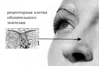
The mucous membrane of the olfactory region is yellow, in contrast to the rest of the bright red part of the mucous membrane of the wall of the nasal cavity. The nasal cavity, like the cavity of the upper respiratory tract, is lined with pseudostratified columnar ciliated epithelium with goblet cells. The epithelium of the olfactory zone occupies the area of the upper nasal passage and the posterosuperior part of the septum and is pseudo-multilayered cylindrical ciliated. It consists of three types of cells: olfactory nerve, basal and supporting. Cell nuclei are arranged in rows. On the surface there is a single row of supporting cells with oval nuclei. Beneath it are numerous rows of olfactory and basal cell nuclei with rounded nuclei.
OLfactory cells.
These are bipolar ganglion cells. Deep in the cell there is a rounded nucleus. In the apical part there is a modified dendrite, which extends to the surface of the mucous membrane and ends in olfactory vesicles equipped with immobile cilia. The cell tapers towards the base, forming an unmyelized axon, which enters the underlying connective tissue and connects with neighboring axons to form bundles of olfactory nerve fibers. The renewal of olfactory cells occurs every 40 days due to neurons that are formed from basal cells. This is the only known case of the formation of new neurons in the postnatal period.
Among all the senses, vision and hearing play the most important and significant role in human life. Therefore, for a long time, it was these channels that connect us with the outside world that were studied most actively. But the olfactory analyzer attracted the attention of physiologists to a much lesser extent. Indeed, the sense of smell in humans, and in primates in general, is relatively poorly developed. And yet, its role in our lives should not be underestimated.
Even a newborn baby reacts to odorous substances from the first hours of life, and at the 7th–8th month of life he develops conditioned reflexes to “pleasant” and “unpleasant” odors.
A person can perceive more than 10,000 odors. Some of them can excite or discourage appetite, change mood and desires, increase or decrease performance, and even force you to buy something you don’t really need. In many stores in Europe and America, fragrances are used with all their might to attract customers. According to an American marketing service, simply aromatizing the air in a store can increase sales by 15%. There are even five scents that, when present in the store, can “provoke” a visitor to buy underwear and outerwear. These are vanilla, lemon, mint, basil and lavender. Grocery supermarkets should be filled with fresh smells: warm bread, cucumbers and watermelons. There are also holiday smells. For example, before the New Year, stores should smell like tangerines, cinnamon and spruce or pine needles. For most people, these smells are strongly associated with memories of the holiday and give them pleasure. However, in some people (especially children), sprayed aromatic substances can cause allergies. So, maybe it’s good that “advertising” fragrances are not sprayed in our stores yet.
 Smells can easily “stir up” our memory and bring back long-forgotten sensations, for example from childhood. The fact is that the centers of the olfactory analyzer are located in humans in the ancient and old cerebral cortex. Next to the olfactory center is the center responsible for our emotions and memory. Therefore, smells are emotionally charged for us, awakening not logical, but emotional memory.
The perception of smell by our olfactory system begins with the nose, or more precisely, with the olfactory epithelium, located in humans in the upper parts of the middle turbinate, in the superior turbinate and the upper part of the nasal septum. The peripheral processes of the receptor cells of the olfactory epithelium end in an olfactory club, decorated with a bunch of microvilli. It is the membrane of these villi (cilia and microvilli) that is the site of interaction between the olfactory cell and the molecules of odorous substances. In humans, the number of olfactory cells reaches 6 million (3 million in each nostril). This is a lot, but in those mammals in whose life the sense of smell plays a significant role, these cells are immeasurably more numerous. For example, a rabbit has about 100 million of them!
In the human embryo, the development of olfactory cells occurs quite quickly. Already in an 11-week fetus they are well differentiated and presumably capable of performing their function.
The receptor cells of the olfactory epithelium are constantly renewed. The life of one cell lasts only a few months or even less. When the olfactory epithelium is damaged, cell regeneration is significantly accelerated.
But how does the excitation of olfactory cells occur? In the last decade, it has become clear that the main role in this process belongs to receptor proteins, whose molecules, interacting with molecules of odorant substances, change their conformation. This leads to the launch of a whole chain of complex reactions, as a result of which the sensory signal is converted into a universal signal from nerve cells. Next, from the receptor cells along their axons, which form the olfactory nerve, the signal is transmitted to the olfactory bulbs. Here its primary processing takes place, and then the signal travels along the olfactory nerve to the brain, where its final analysis occurs.
The ability to perceive odors changes as a person ages. Olfactory acuity reaches its maximum by age 20, stays at the same level for about 30–40 years, and then begins to decline. A particularly noticeable decrease in the acuity of smell occurs in people over 70 years of age, and sometimes even 60 years of age. This phenomenon is called senile hyposmia, or presbiosmia, and is not nearly as harmless as it might seem. Older people gradually cease to perceive the smell of food and therefore lose their appetite. After all, the aroma of food is one of the necessary conditions for the production of digestive juices in the gastrointestinal tract. No wonder they say: “... such a wonderful smell that even my mouth started watering...”. In addition, taste and olfactory perceptions are very close. The odorous substances contained in food products enter the nasal cavity through the nasopharynx, and we smell their aroma. But when we have a runny nose, no matter what we eat, it feels like we are chewing tasteless cardboard. Elderly people with a sharply reduced sense of smell perceive food in the same way. They also lose the ability to determine the quality of food by smell, and therefore can become poisoned by eating low-quality food. It also turns out that older people no longer perceive the smell of mercaptans as unpleasant. Mercaptans are substances added to natural gas used in everyday life (which itself does not smell like anything from a human point of view) specifically so that its leak can be detected by smell. Old people stop noticing this smell...
But even among young people, sensitivity to the smell of the same substances varies greatly. It also changes depending on environmental factors (temperature, humidity), emotional state and hormonal levels. In pregnant women, for example, against the background of a general decrease in the acuity of smell, sensitivity to certain odors sharply increases. In general, the range of threshold concentrations of various odorous substances perceived by humans is very large - from 10-14 to 10-5 mol per 1 liter of air.
Until now, we have talked mainly about external odors that come from the world around us. But among the odorous substances, there are also those that are released by our body itself and are capable of causing certain behavioral and physiological reactions in other people. Substances with such properties are called pheromones. In the animal world, pheromones play a huge role in the regulation of behavior - we have already written about this in our newspaper (No. 10/1996 and No. 16/1998). Substances have also been discovered in humans that have a certain pheromonal effect during our communication. Such substances are found, for example, in human sweat. In the 70s XX century researcher Martha McClintock discovered that women who live in the same room (for example, in a dormitory) for a long time synchronize their menstrual cycles. And the smell of the secretion of male sweat glands causes the normalization of unstable menstrual cycles in women.

Tapestry “Lady with a Unicorn” – an allegorical depiction of the sense of smell
The smell of the secretion secreted by our axillary sweat glands depends both on the substances secreted by the body itself and on the bacteria present in the sweat glands. After all, it is known that fresh axillary sweat itself (produced profusely, for example, in hot weather) does not have a strong specific odor. But the activity of bacteria contributes to the release of odorous molecules, initially associated with special carrier proteins from the group of lipocaines.
The chemical composition of men's and women's sweat differs greatly. In women, it is associated with the phases of the menstrual cycle, and a man who has been in an intimate relationship with a woman for a long time is able to determine the time of ovulation in his partner by smell. True, as a rule, this happens unconsciously - it’s just that during this period the girlfriend’s smell becomes the most attractive to him.
In the secretions of the sweat glands of both men and women, in addition to other components, two odorous steroids are present - androstenone (ketone) and androstenol (alcohol). For the first time, these substances were identified as components of the sex pheromone contained in boar saliva. Androstenone has a strong, specific odor, which for many people is similar to the smell of urine. The smell of androstenol is perceived as musky or sandalwood. The content of androstenone and androstenol in men's axillary sweat is much higher than in women's. Studies have shown that the smell of androstenone can affect the physiological and emotional state of people, in particular, suppress the effect of synchronization of sexual cycles described above in women living in the same room. In some situations, the faint smell of androstenone creates a comfortable state of “security” in women, while in men, on the contrary, it causes discomfort and is associated with competition and aggression.
Representatives of different cultures may perceive the same odors differently. Such differences were revealed in a completely unique survey conducted in 1986 by National Geographic magazine. The next issue of this magazine included samples of six odorous substances: androstenone, isoamyl acetate (smells like pear essence), galaxolide (smells like synthetic musk), eugenol, a mixture of mercaptans and rose oil. The substances were enclosed in microcapsules applied to paper. When the paper was rubbed with a finger, the capsules were easily destroyed and the smell was released. Readers were asked to smell the proposed substances and then answer the questionnaire. It was necessary to evaluate the intensity of the proposed odors, define them as pleasant, unpleasant or neutral, and talk about the emotions and memories they evoked. Respondents were also asked to indicate their age, gender, occupation, country of residence, race, presence of diseases, etc. For women, it was necessary to indicate the presence of pregnancy. Letters with completed questionnaires came from more than 1.5 million people living on different continents!

Baker of the House of Amun donating incense to Osiris
Many of those who responded did not smell androstenone at all, and the number of people who were not sensitive to this smell varied greatly in different regions of the globe. So, if in the USA about 30% of women did not feel this smell, then among white women living in Africa there were half as many - about 15%.
We have already talked about the loss of olfactory acuity in older people, which was also clearly revealed during this study. The survey also confirmed that smokers have a much worse sense of smell than non-smokers.
People who, for various reasons, had completely lost their sense of smell also sent their answers to National Geographic. It turns out that there are a lot of such people, including among young people. According to the US National Institutes of Health, in 1969, smell disorders were noted in 2 million people, and by 1981 this figure had increased to 16 million! This situation is largely due to the deterioration of the environmental situation. Among the patients at the Smell and Taste Clinic in Washington, 33% of patients with dysosmia (impaired sense of smell) are people aged 17–20 years. According to researcher Hendricks, in 1988, 1% of the Dutch population had problems with their sense of smell. As for our country, very often people, overwhelmed by other problems, simply do not pay attention to such a “trifle” as a violation or lack of sense of smell. And if they do, they don’t know whether medical help is possible in this case and where to go for it. Treatment of people with impaired sense of smell is carried out in Moscow, at the ENT clinic of the Moscow Medical Academy. THEM. Sechenov.
What can cause a violation of the sense of smell? Most often, the corresponding disorders are associated with damage to the receptor apparatus of the olfactory analyzer (about 90% of cases), with damage to the olfactory nerve - about 5% of cases, and with damage to the central parts of the brain - the remaining 5% of cases.
The causes of olfactory disorder at the “receptor level” are very diverse and numerous. These include injuries to the olfactory zone and cribriform plate, and inflammatory processes in the nasal cavity, and traumatic brain injuries, and drug intoxication, and allergic reactions, and mutations, and vitamin deficiencies (for vitamins A and B12), and intoxication with heavy metal salts (cadmium , mercury, lead), and inhalation of vapors of irritating substances (formaldehyde), and viral infections (mainly the influenza virus), and ionizing radiation, and much more.
The causes of damage to the olfactory nerve are most often due to infectious diseases, metabolic disorders, toxic effects of drugs, nerve damage during surgery and tumors.
Damage to the centers of the olfactory analyzer can be caused by traumatic brain injury, cerebrovascular accident, brain tumors, genetic and infectious diseases, demyelinating processes, Parkinson's disease, Alzheimer's disease. In the latter two diseases, a decrease in the acuity of smell is often detected in the early stages, which allows treatment to begin earlier.
What is a violation of the sense of smell? This may be a complete lack of ability to perceive smells (anosmia) or a decrease in the acuity of smell (hyposmia) of varying degrees. A violation of the sense of smell can also be expressed in the form of a distortion in the perception of odors (aliosmia), in which all odors are perceived “in the same manner.” For example, with cacosmia, all odors seem putrid and fecal; with torsosmia - chemical, bitter, burning or metal odors; with parosmia, “garlic smells like violets.” Mixed cases and phantosmia – olfactory hallucinations – are also possible.
Many of the described smell disorders can be successfully treated, especially if you do not delay visiting a doctor.
Olfactory organ is a chemoreceptor. It perceives the action of odorant molecules. This is the most ancient type of reception. The olfactory analyzer consists of three parts: the olfactory region of the nasal cavity (peripheral part), the olfactory bulb (intermediate part), and also the olfactory centers in the cerebral cortex.
Development of the sense of smell. The source of formation of all parts of the olfactory organ is the neural tube, symmetrical local thickenings of the ectoderm - olfactory placodes located in the area of the anterior part of the head of the embryo and mesenchyme. The placode material is invaginated into the underlying mesenchyme, forming olfactory sacs connected to the external environment through openings (future nostrils). The wall of the olfactory sac contains stem cells, which in the 4th month of embryogenesis, through divergent differentiation, develop into neurosensory (olfactory) cells that support basal epithelial cells. Some of the cells of the olfactory sac go to build the olfactory (Bowman's) gland.
At the base of the nasal septum The vomeronasal (Jacobson) organ is formed, the neurosensory cells of which respond to pheromones.
Structure of the sense of smell. The olfactory lining of the peripheral part of the olfactory analyzer is located on the superior and partially middle concha of the nasal cavity. Its total area is about 10 cm2. The olfactory region has an epithelial-like structure. The receptor part of the olfactory analyzer is delimited from the underlying connective tissue by the basement membrane. Olfactory neurosensory cells are spindle-shaped with two processes. Based on their shape, they are divided into rod-shaped and cone-shaped. The total number of olfactory cells in humans reaches 400 million, with a significant predominance of rod-shaped cells.
Peripheral process The olfactory neurosensory cell is 15-20 microns long and has a thickening at the end called the olfactory club. On the rounded top of the olfactory clubs there are 10-12 olfactory hairs - antennae. Their length reaches 2-3 microns. The antennae have an ultrastructure characteristic of cilia, i.e. they contain 9 peripheral and 2 central paired protofibrils extending from typical basal bodies. The antennas perform continuous automatic pendulum-type movements. The top of the antennas moves along a complex trajectory, thereby increasing the possibility of their contact with molecules of odorous substances. The antennae are immersed in a liquid medium, which is the secretion of the tubular-alveolar olfactory glands (Bowman's). They are characterized by a merocrine type of secretion. The secretion of these glands moisturizes the surface of the olfactory lining.
Central process olfactory neurosensory cell - axon, is sent to the intermediate part of the olfactory organ - the olfactory bulb and establishes a synaptic connection there in the form of a glomerulus with mitral neurons. The following layers are distinguished in the olfactory bulb: 1) layer of olfactory glomeruli, 2) outer granular layer, 3) molecular layer, 4) layer of mitral cells, 5) inner granular layer, 6) layer of centrifugal fibers.
Central part of the olfactory organ is localized in the hippocampus and in the hippocampal gyrus of the cerebral cortex, where the axons of mitral cells are sent and form synaptic connections with neurons.
Thus, the organ sense of smell(olfactory region of the nasal cavity and olfactory bulb), like the organ of vision, has a layered arrangement of neurons, which is characteristic of screen nerve centers.
Supporters epithelial cells of the olfactory region- highly prismatic cells with microvilli, located in the form of a multirow epithelial layer, providing spatial organization of neurosensory cells. Some of these cells are secretory and also have phagocytic ability. Cuboidal basal epithelial cells are poorly differentiated (cambial) and serve as the source of the formation of new cells of the olfactory lining. The cycle of physiological regeneration of neurosensory cells is estimated at 30-35 days.
Bowman's glands consist of secretory and myoepithelial cells. The secretion of the glands dissolves odorous substances that interact with the receptor-transductor system of the cilia of the neurosensory cell. This causes a change in membrane potential, which is transmitted through a chain of neurons to the central part of the olfactory organ.


