Main organs of the immune system. Description and principle of operation of the human immune system
Immune system, consisting of special proteins, tissues and organs, daily protects humans from pathogenic microorganisms, and also prevents the influence of some special factors (for example, allergens).
In most cases, she performs a huge amount of work aimed at maintaining health and preventing the development of infection.
Photo 1. The immune system is a trap for harmful microbes. Source: Flickr (Heather Butler)
What is the immune system
The immune system is a special protective system of the body that prevents the effects of foreign agents (antigens). Through a series of steps called the immune response, it “attacks” all microorganisms and substances that invade organ systems and tissues and are capable of causing disease.
Immune system organs
The immune system is amazingly complex. It is able to recognize and remember millions of different antigens, promptly producing the necessary components to destroy the “enemy”.
She includes central and peripheral organs, as well as special cells, which are produced in them and are directly involved in human protection.
Central authorities
The central organs of the immune system are responsible for the maturation, growth and development of immunocompetent cells - lymphopoiesis.
Central authorities include:
- Bone marrow- spongy tissue of a predominantly yellowish hue, located inside the bone cavity. Bone marrow contains immature, or stem cells, which are capable of turning into any, including immunocompetent, cell of the body.
- Thymus gland(thymus). It is a small organ located in the upper part of the chest behind the sternum. In shape, this organ is somewhat reminiscent of thyme, or thyme, the Latin name of which gave the name to the organ. The thymus is primarily where T cells of the immune system mature, but the thymus gland is also capable of inducing or maintaining the production of antibodies against antigens.
- During the prenatal period, the central organs of the immune system also include the liver..
This is interesting! The largest size of the thymus gland is observed in newborns; With age, the organ shrinks and is replaced by fatty tissue.
Peripheral organs
Peripheral organs are distinguished by the fact that they contain mature cells of the immune system that interact with each other and other cells and substances.
Peripheral organs are represented by:
- Spleen. The largest lymphatic organ in the body, located under the ribs on the left side of the abdomen, above the stomach. The spleen contains primarily white blood cells and also helps get rid of old and damaged blood cells.
- Lymph nodes(LNs) are small, bean-shaped structures that house cells of the immune system. The lymph node also produces lymph, a special clear liquid through which immune cells are delivered to various parts of the body. As the body fights an infection, the lymph nodes may increase in size and become painful.
- Clusters of lymphoid tissue, containing immune cells and located under the mucous membranes of the digestive and genitourinary tract, as well as in the respiratory system.
Immune system cells
The main cells of the immune system are white blood cells, which circulate in the body through lymphatic and blood vessels.
The main types of leukocytes capable of an immune response are the following cells:
- Lymphocytes, which allow you to recognize, remember and destroy all antigens that invade the body.
- Phagocytes, absorbing foreign particles.
Phagocytes can be various cells; the most common type are neutrophils, which primarily fight bacterial infection.
Lymphocytes are located in the bone marrow and are represented by B cells; If lymphocytes are found in the thymus, they mature into T-lymphocytes. B and T cells have different functions:
- B lymphocytes try to detect foreign particles and send a signal to other cells when an infection is detected.
- T lymphocytes destroy pathogenic components identified by B cells.
How the immune system works
When antigens (that is, foreign particles that invade the body) are detected, they are induced B lymphocytes, producing antibodies(AT) are specialized proteins that block specific antigens.
Antibodies are able to recognize the antigen, but cannot destroy it on their own - this function belongs to T cells, which perform several functions. T cells can not only destroy foreign particles (there are special T-killers, or “killers” for this), but also participate in the transmission of the immune signal to other cells (for example, phagocytes).
Antibodies, in addition to identifying antigens, neutralize toxins produced by pathogenic organisms; also activate complement - part of the immune system that helps destroy bacteria, viruses and other and foreign substances.
Recognition process
After antibodies are formed, they remain in the human body. If the immune system encounters the same antigen in the future, infection may not develop: for example, after suffering from chickenpox, a person no longer gets sick from it.
This process of recognizing a foreign substance is called antigen presentation. The formation of antibodies during re-infection is no longer required: the destruction of the antigen by the immune system is carried out almost instantly.
Allergic reactions
Allergies follow a similar mechanism; A simplified diagram of the development of the state is as follows:
- Primary entry of the allergen into the body; It is not expressed clinically in any way.
- Antibody formation and fixation on mast cells.
- Sensitization - increased sensitivity to an allergen.
- Re-entry of the allergen into the body.
- Release of special substances (mediators) from mast cells with the development of a chain reaction. Subsequent produced substances affect organs and tissues, which is determined by the appearance of symptoms of the allergic process.
 Photo 2. An allergy occurs when the body’s immune system mistakes a substance as harmful.
 Immune system unites organs and tissues whose function is to protect the body from genetically foreign substances coming from outside or formed in the body itself. The organs of the immune system produce immunocompetent cells (lymphocytes, plasma cells), biologically active substances (antibodies) that recognize and destroy cells and other foreign substances (antigens) that have entered the body or formed in it.
The immune system includes all organs that are built from lymphoid tissue and carry out protective reactions in the body, create immunity-immunity to foreign antigenic substances.
The organs of the immune system include red bone marrow, thymus, tonsils, appendix, lymph nodes, spleen, accumulation of lymphoid tissue (lymphoid nodules) in the walls of the hollow internal organs of the digestive, respiratory systems and genitourinary apparatus (Fig. 360).
The bone marrow and thymus are the central organs of the immune system, where lymphocytes are formed from bone marrow stem cells. In the bone marrow, B lymphocytes are formed from its stem cells. In the thymus, differentiation of T-lymphocytes (thymus-dependent) occurs. B-lymphocytes and T-lymphocytes from the bone marrow and from the thymus through the bloodstream enter the peripheral organs of the immune system, which include tonsils, lymphoid (Peyr's) patches, appendix, single lymphoid nodules, lymph nodes and spleen.
Central authorities immune systems are located in the human body in well-protected places (bone marrow - in the bone marrow cavities, thymus - in the chest cavity, behind the manubrium of the sternum). Peripheral organs immune systems are located in places of possible penetration of foreign substances into the body or along the paths of their movement in the body itself. The tonsils are located in the walls of the initial section of the digestive tube and respiratory tract, on the border between the oral cavity, nose and the cavity of the pharynx and larynx. Lymphoid (Peyre's) plaques are located in the walls of the small intestine (mainly the ileum), the appendix is near the cecum, with a particularly abundant microflora.
In the mucous membrane of the digestive, respiratory and urinary tract organs there are numerous single lymphoid nodules that perform immune surveillance functions at the border of the body and the external environment (inhaled air, contents of the digestive tract). Lymph nodes, which are biological filters, lie on the paths of lymph flow (tissue fluid) from organs and tissues into the venous system. Particles of dead cells and coarse proteins, together with tissue fluid, enter the lymphatic channel, are retained and neutralized in the lymph nodes. The spleen, whose function is the immune control of blood, is located on the path of its flow from the arterial system to the portal vein.
Diffuse lymphoid tissue, represented by individual scattered cells of the lymphoid series, in some places forming not very dense cell clusters, is present in those organs where the antigenic danger is not very great. In places of constant antigenic potential
Rice. 360.Diagram of the location of the central and peripheral organs of the immune system in the human body.
1 - red bone marrow, 2 - thymus, 3 - lingual tonsil, 4 - palatine tonsil, 5 - tubal tonsil, 6 - pharyngeal tonsil, 7 - lymphoid nodules in the walls of the trachea and bronchi, 8 - lymph nodes (axillary), 9 - spleen, 10 - lymphoid nodules of the appendix, 11 - lymphoid nodules in the walls of the colon.
actions (tonsils, mucous membrane of the stomach, intestines, lymph nodes, spleen), lymphocytes form dense clusters measuring 0.5-1 mm, called lymphoid nodules, with reproduction centers (germentative centers).
Bone marrow(medulla ossium) is a hematopoietic organ and the central organ of the immune system. There are red bone marrow, which in an adult is located in the cells of the spongy substance of flat and short bones, the epiphyses of long (tubular) bones, and yellow bone marrow, which fills the bone marrow cavities of the diaphyses of long (tubular) bones. Red bone marrow contains hematopoietic stem cells - the precursors of all blood cells and the immune system (lymphocytes).
Thymus
Thymus(thymus), which was formerly called the thymus gland, is the central organ of immunogenesis. In the thymus, from stem cells coming here from the bone marrow with the blood flow, T-lymphocytes are formed, which leave the thymus with the blood flow and populate the thymus-dependent zones of the peripheral organs of immunogenesis. The thymus also secretes substances that affect the functions of T-lymphocytes.
The thymus consists of two asymmetrical right and left lobes, which are fused to each other at the level of their middle.
The thymus has a thin connective tissue capsule. The thymic parenchyma consists of darker cortex(cortex thymi) and lighter medulla(medulla thymi), occupying the central part of the thymus lobules. In the loops of the network formed by reticular fibers and cells, there are thymic lymphocytes(thymocytes), which lie more densely in the cortex than in the medulla, and stellate-shaped multi-processed epithelial cells - epithelioreticulocytes. The medulla also contains thymic corpuscles(corpuscula thymici), Hassal's bodies, formed by concentrically lying, highly flattened epithelial cells.
Innervation of the thymus: branches of the right and left vagus nerves, as well as branches of the cervicothoracic (stellate) and superior thoracic nodes of the sympathetic trunk.
Blood supply:branches of the internal mammary artery. Vienna The thymus drains into the brachiocephalic and internal mammary veins.
Tonsils
Tonsils:lingual and pharyngeal (unpaired), palatine and tubal (paired) - located in the area of the root of the tongue, pharynx and nasal part of the pharynx, respectively. They are diffuse accumulations of lymphoid tissue containing small, denser cell masses - lymphoid nodules.
Lingual tonsil (tonsilla lingualis) unpaired, located under the multilayered epithelium of the mucous membrane of the root of the tongue, often in the form of two accumulations of lymphoid tissue.
The surface of the tongue above the tonsil is lumpy, between the tubercles there are openings of the mucous glands located in the thickness of the root of the tongue.
The lingual tonsil reaches its largest size by the age of 14-20; its length is 18-25 mm and its width is 18-25 mm. The lingual tonsil does not have a capsule.
The lingual tonsil consists of lymphoid nodules, the number of which (80-90) is greatest in childhood, adolescence and young adulthood.
Innervation of the lingual tonsil: branches of the glossopharyngeal and vagus nerves, as well as sympathetic fibers of the external carotid plexus.
Blood supply:branches of the right and left lingual arteries. Venous blood drains into the lingual vein.
Pharyngeal tonsil (tonsilla pharyngealis), unpaired, is located in the vault of the pharynx, where diffuse lymphoid tissue and lymphoid nodules are located, mainly with reproductive centers.
Innervation:branches of the facial, glossopharyngeal, vagus nerves and sympathetic periarterial plexuses.
Blood supply:branches of the ascending pharyngeal arteries. Venous blood
Palatine tonsil (tonsilla palatina) steam room, located in the tonsillar fossa between the palatoglossal and velopharyngeal arches. The medial (free) surface of the tonsil faces the pharynx. On this surface there are almond dimples into which almond crypts open. In the thickness of the tonsil, along its crypts, there are lymphoid nodules, mainly with reproduction centers. Around the lymphoid nodules there is diffuse lymphoid tissue (Fig. 361).
Innervation:branches of the greater palatine nerve (from the pterygopalatine ganglion), the tonsil branch of the glossopharyngeal nerve and sympathetic fibers from the internal carotid plexus.
Blood supply:branches of the lingual, ascending pharyngeal and descending palatine arteries. Venous blood flows into the veins of the pterygoid plexus.
Tubal tonsil (tonsilla tubaria) steam room, located in the area of the tubal ridge, near the pharyngeal opening of the auditory tube. The tonsil consists of diffuse lymphoid tissue and a few lymphoid nodules.
Innervation:branches of the facial, glossopharyngeal and vagus nerves and periarterial sympathetic plexuses.
Blood supply:branches of the ascending pharyngeal artery. Venous blood flows into the veins of the pharyngeal plexus.
Appendix
Appendix (appendix, appendix vermiformis) extends from the lower part of the cecum, has numerous lymphoid nodules in its walls and internodular lymphoid tissue between them. The number of lymphoid nodules in the walls of the appendix in children and adolescents reaches 800, the nodules are located one above the other in 2-3 rows.
Innervation:fibers of the vagus nerves and the celiac (sympathetic) plexus.

Rice. 361.Microscopic structure of the palatine tonsil.
1 - crypts of the tonsil, 2 - integumentary epithelium, 3 - lymphoid nodules of the tonsil.
Blood supply:cecum branches of the ileocolic artery. Venous blood flows into the vein of the same name.
Lymphoid plaques of the small intestine
Lymphoid plaques (noduli lymphoidei aggregati), or group lymphoid nodules (Peyer's patches) are a cluster of lymphoid nodules located in the walls of the small intestine, mainly in its final section (Fig. 362). Lymphoid plaques look like oval or round formations, slightly protruding into the intestinal lumen. One plaque has from 5 to 150 or more lymphoid nodules, between which diffuse lymphoid tissue is located.
Single lymphoid nodules
Single lymphoid nodules (noduli lymphoidei solitarii) are present in the mucous membrane and submucosa of all tubular organs of the digestive, respiratory systems and genitourinary apparatus. Lymphoid nodules are located at different distances from each other and at different depths. Often the nodules lie so close to the epithelial cover that the mucous

Rice. 362.Group and single lymphoid nodules in the wall of the small intestine.
1 - serous membrane, 2 - muscular membrane, 3 - mucous membrane, 4 - mesentery of the small intestine, 5 - single lymphoid nodules, 6 - group lymphoid nodule (Peyer's patch), 7 - circular folds of the mucous membrane.
The melted shell rises above them in the form of small mounds. In the small intestine in childhood, the number of nodules varies from 1200 to 11000, in the large intestine - from 2000 to 9000, in the walls of the trachea - from 100 to 180, in the bladder - from 80 to 530. Diffuse lymphoid tissue is also present in the mucous membrane of all organs of the digestive, respiratory systems and genitourinary apparatus.
Innervationlymphoid nodules and lymphoid plaques are carried out along the branches of the vagus nerves and the celiac plexus.
Blood supply:peri-nodular hemocapillary networks formed by branches of organ arteries. Venous blood flows into the veins of the same name.
Spleen
Spleen(lien, splen), which carries out immune control of the blood, is located in the left hypochondrium, at the level of 9-11 ribs. The spleen has diaphragmatic and visceral surfaces. Diaphragmatic surface(facies diaphragmatica) facing the diaphragm. Anteromedial (visceral) surface(facies visceralis) contains the gate of the spleen, through which the artery and nerves enter the organ and the vein exits.
The spleen is covered on all sides by peritoneum, under which there is a thin fibrous membrane. Connective tissue trabeculae extend from the fibrous membrane into the organ, between which there is parenchyma, or pulp (pulp), spleen(pulpa splenica). Highlight red pulp(pulpa rubra), located between the venous vessels - the sinuses of the spleen, consisting of loops of reticular tissue filled with erythrocytes, leukocytes, lymphocytes, macrophages, and white pulp(pulpa alba), formed by periarterial lymphoid couplings, lymphoid nodules and macrophage-lymphoid couplings (ellipsoids), consisting of lymphocytes and other cells of lymphoid tissue (Fig. 363).
Periarterial lymphoid couplings in the form of several layers of lymphoid cells surround the pulp arteries along their entire length. Lymphoid nodules form in the thickness of the periarterial lymphoid couplings. Around the arterioles and capillaries there are 2-3 layers of cells of the lymphoid series - macrophage-lymphoid couplings (ellipsoids), which have a spindle shape.
Innervation of the spleen: sympathetic fibers from the celiac plexus and branches of the vagus nerves.
Blood supply:splenic artery. Venous blood flows through the splenic vein.
Lymph nodes
Lymph nodes (nodi lymphatici) are located on the paths of lymph flow from organs and tissues to the lymphatic ducts and lymphatic trunks flowing into large veins in the lower parts of the neck. Lymph nodes are biological filters for tissue fluid and the particles of cells contained in it that have died as a result of cellular renewal, and other foreign substances of endogenous and exogenous origin. Lymph flowing through the sinuses of the lymph nodes is filtered through loops of reticular tissue. The lymph receives lymphocytes formed in the lymphoid tissue of these lymph nodes. Lymph nodes are usually located in groups. Groups of lymph nodes are named by the area of their location: (inguinal, lumbar, etc.) or by the name of the blood vessel next to which they are located (celiac, iliac lymph nodes). Lymph nodes adjacent to the walls of cavities are called parietal, parietal lymph nodes(nodi lymphatici parietales), located near the internal organs - visceral lymph nodes(nodi lymphatici

Rice. 363.Diagram of the location of white pulp in the parenchyma of the spleen.
1 - fibrous membrane, 2 - trabecula of the spleen, 3 - venous sinuses, 4 - ellipsoid arteriole (ellipsoid), 5 - brush arterioles, 6 - central artery, 7 - lymphoid nodule, 8 - lymphoid periarterial coupling, 9 - red pulp , 10 - pulp artery, 11 - splenic vein, 12 - splenic artery, 13 - trabecular artery and vein.
viscerales). There are superficial and deep lymph nodes. The shape of the lymph nodes is very different.
On the outside, the lymph node is covered with a connective tissue capsule, from which capsular trabeculae extend into the organ. At the point where the lymphatic vessels exit the lymph node, there is a slight depression - gates(hilus), in the area of which the capsule thickens, forms a portal (hilar) thickening (Fig. 364). Portal (hilar) trabeculae extend from the portal thickening into the node. Through the gate, an artery, nerves enter the lymph node, veins and efferent lymphatic vessels exit. Between the trabeculae of the lymph node there are reticular fibers that form a network in the loops of which lymphoid tissue is located. The parenchyma of the lymph node is divided into cortex and medulla. Cortex(cortex) darker, occupies the peripheral parts of the node. Lighter medulla(medulla) lies closer to the gate of the lymph node. The cortex contains lymphoid nodules with and without a reproductive center. Around the lymphoid nodules there is diffuse lymphoid tissue, in which an internodular zone is distinguished - the cortical plateau. Inward from the lymphoid nodules, at the border with the medulla, there is a strip of lymphoid tissue called pericortical

Rice. 364.Microscopic structure of the lymph node.
1 - capsule, 2 - trabecula, 3 - afferent lymphatic vessel, 4 - subcapsular lymphatic sinus, 5 - cortical substance, 6 - poracortical (thymus-dependent) zone, 7 - lymphoid nodule, 8 - proliferation center of the lymphoid nodule, 9 - cortical lymphatic sinus , 10 - pulpal cords, 11 - cerebral sinuses, 12 - portal sinuses, 13 - efferent lymphatic vessel, 14 - portal thickening, 15 - blood vessels.
(paracortical) substance (paracortex), where there are mainly T-lymphocytes, as well as post-capillary venules. Through the walls of the venules, lymphocytes migrate into the bloodstream from the parenchyma of the lymph node and back. The medulla is formed by strands of lymphoid tissue - pulpy cords(chordae medullares), going from the cortex to the gate of the lymph node. Together with the lymphoid nodules, the pulp cords form the B-dependent zone.
lymphatic sinuses (sinus marginalis) to portal sinus(sinus hilaris). Along the capsular trabeculae lie cortical sinuses(sinus corticalis), along the pulpy cords are the sinuses of the medulla (sinus medullaris), which reach the gates of the lymphatic channel. Near the portal thickening, the sinuses of the medulla flow into the portal sinus located here. In the sinuses there is a fine-mesh network formed by reticular fibers and cells.
The parenchyma of the lymph node is penetrated by a dense network of narrow slits - lymphatic sinuses(sinus lymphaticus), through which the lymph entering the node flows from subcapsular (marginal) sinus(sinus marginalis) to portal sinus(sinus hilaris). Along the capsular trabeculae lie
cortical sinuses (sinus corticalis), along the pulpy cords - medullary sinuses(sinus medullaris), which reach the gates of the lymphatic bed. Near the portal thickening, the sinuses of the medulla flow into the portal sinus located here. In the sinuses there is a fine-mesh network formed by reticular fibers and cells.
Lymphatic system
Lymph nodes, lymphatic capillaries and vessels, ducts and trunks through which lymph flows are united under the general name - lymphatic system(systema lymphaticum) (Fig. 365).
Lymphatic capillaries (vasa lymphocapillaria) are the initial link of the lymphatic system. Tissue fluid, together with the substances it contains (large protein molecules, particles of dead cells, tumor cells), including foreign particles, is absorbed into the lumen of the lymphatic capillaries and is called lymph(lymph). Lymphatic capillaries are present in all organs and tissues of the human body, except for the brain and spinal cord, eyeball, inner ear, epithelial skin and mucous membranes, cartilage, splenic parenchyma, bone marrow and placenta. The diameter of lymphatic capillaries varies from 10 to 200 microns. When connected to each other, capillaries form in organs and tissues closed lymphocapillary networks(rete lymphocapillaria). The walls of lymphatic capillaries are built from a single layer of endothelial cells.
Lymphatic vessels (vasa lymphatica) are formed by the fusion of lymphatic capillaries. The walls of the lymphatic vessels are thicker, they consist of three layers (inner shell- tunica intima, middle shell- tunica media and outer shell- tunica externa). Lymphatic vessels have valves, the presence of which gives these vessels a characteristic bead-shaped appearance. The valves of the lymphatic vessels, formed by the folds of the inner membrane, allow lymph to pass in one direction - from the place of its formation in the capillaries towards the lymph nodes. From the lymph nodes, through their efferent lymphatic vessels, lymph flows either to the next (along the lymph flow) lymph nodes, or to the collector vessels - lymphatic trunks and lymphatic ducts, which flow into the venous angle formed on the right and left at the connection of the internal jugular and subclavian veins of the corresponding sides
Rice. 365.Human lymphatic system. Front view.
1 - lymphatic vessels of the face, 2 - submandibular lymph nodes, 3 - mental lymph nodes, 4 - mouth of the thoracic duct, 5 - anterior mediastinal lymph nodes, 6 - axillary lymph nodes, 7 - superficial ulnar lymph node, 8 - superficial lymphatic vessels of the forearm , 9 - lumbar lymph nodes, 10 - subaortic lymph node, 11 - common iliac lymph nodes, 12 - superficial inguinal lymph nodes, 13 - medial group of superficial lymphatic vessels of the leg, 14 - lateral group of superficial lymphatic vessels of the leg, 15 - superficial lymphatic vessels feet, 16 - deep lymphatic vessels of the foot, 17 - deep lymphatic vessels of the leg, 18 - deep lymphatic vessels of the thigh, 19 - deep lymphatic vessels of the palm, 20 - deep inguinal lymph nodes, 21 - external and internal iliac lymph nodes, 22 - deep lymphatics vessels of the forearm, 23 - thoracic duct, 24 - deep ulnar lymph node, 25 - intercostal lymph nodes, 26 - subclavian trunk, 27 - jugular trunk, 28 - deep cervical lymph nodes, 29 - jugular-digastric lymph node, 30 - mastoid lymph nodes nodes, 31 - preauricular lymph nodes.

Lymphatic trunks (trunci lymphatici) and lymphatic ducts(ductus lymphatici) are large lymphatic vessels that collect lymph (tissue fluid) from large parts of the body. There are six large lymphatic ducts and trunks in the human body. The thoracic duct, left jugular and left subclavian trunks flow into the left venous angle) into the right venous angle - the right lymphatic duct, right jugular and right subclavian trunks.
IN right subclavian trunk(truncus subclavius dexter) lymph flows from the right upper limb, into right jugular trunk(truncus jugularis dexter) - from the right half of the head and neck. IN right lymphatic duct(ductus lymphaticus dexter) the right bronchomediastinal trunk flows into, collecting lymph from the organs of the right half of the thoracic cavity.
Left subclavian trunk (truncus subclavius sinister) collects lymph from the left upper limb, left jugular trunk(truncus jugularis sinister) - from the left half of the head and neck. The largest lymphatic vessel, which also flows into the left venous angle, is thoracic duct(ductus thoracicus), through which lymph flows from the lower extremities, walls and organs of the pelvis and abdominal cavity, as well as the left half of the chest cavity.
The body is constantly under vigilant protection, which protects it from foreign particles. The defense system is immunity. It can be a collection of organs and tissues whose cells neutralize and remove harmful agents. Thanks to this defense system, people can fight diseases. Where is the human immune system located?
There is no clear answer to this question. Immunity is an abstract concept; it is not concentrated in one place, but is scattered throughout the body. Its failure leads to many adverse consequences. A person becomes vulnerable to foreign agents. He cannot fight infections and maintain his health. A number of pathological conditions arise. Depending on the mechanism of operation, immunity can be:
- Cellular;
- Humoral.
Each of them carries out its protective function through special cells. The first type is through T cells or T lymphocytes, which are divided into killer T cells, helper T cells, macrophages and neutrophils. Humoral is carried out thanks to B-lymphocytes and the antibodies they produce.
Another classification of immunity divides it into:
- Nonspecific, otherwise congenital;
- Specific, which is produced throughout life.
And also the protective system can be:
- Natural, developed after an illness;
- Artificial or passive, resulting from medical interventions - vaccination.

Where is
Immunity is a broad concept that combines many systems and tissues involved in the production of special substances. All protection organs are divided into two large groups:
- Central – thymus and bone marrow, which are responsible for the production of lymph cells;
- Peripheral - spleen, lymph nodes, tonsils and groups of lymphoid formations. Their task is differentiation.
It is impossible to say where immunity is located in the human body. This is a general concept that reflects the work of many tissues and systems. The thymus, or otherwise the thymus gland, is the site of formation of cellular immunity, that is, T cells. This organ is located behind the sternum and has age-related characteristics. In children and young people it is actively developed, and involution occurs over the years. This is reflected in a decrease in protective activity among older people.
Another central organ of the immune system is the bone marrow. It is represented by soft spongy tissue, which is located in tubular and flat bones. Its task is the formation of blood cells - leukocytes and erythrocytes, as well as platelets. The bone marrow produces B lymphocytes, which are the weapon of the humoral response.
One of the organs of peripheral immunity is the spleen. Its role is reduced to lymph production, disposal of old and defective red blood cells, as well as storage of blood cells. The spleen is often called a blood depot and a graveyard of red blood cells. It is located in the abdominal area under the left hypochondrium.
Lymph nodes are called biological filters. These are small spherical formations, normally up to 1 cm in size. Lymph nodes follow the course of the arteries. There are submandibular, postauricular, supra- and subclavian, axillary, popliteal, inguinal nodes. They are connected by ducts and together form the lymphatic system. Inflammatory processes are often accompanied by an increase in these nodes. They can reach the size of a chicken egg.
- lymphoid tissue that is located on both sides of the pharynx - these are the tonsils. Another similar island is localized in the intestinal walls and is called Peyer's patches. The exact place of their concentration is the appendix, which is also considered an immune organ. The ducts connecting the lymphoid formations contain lymph, a colorless liquid that includes a large number of defense cells.
Where is immunity located? It permeates the entire human body. Even where there are no protective tissues, there are lymph nodes and ducts. Defense never leaves its post. A person is under the control of a security system 24 hours a day.

How is it produced?
The role of all immune organs comes down to one thing – protective cells. Some form them, others differentiate, and others accumulate, while others act as a reservoir. The most important immune cell products include:
Thanks to the immune organs, the production of these cells is possible. They are direct participants in the battlefield. “Soldiers” of the defense system who fight against strangers who have broken into the body.
Functions
The main task of immunity is defense against unwanted substances. Among the main ones are:
- Preventing the introduction of foreign agents through the presence of biological barriers;
- Destruction of defective and old cells of the body whose life cycle has come to an end;
- Neutralization of a harmful microbe when it enters;
- Elimination, that is, removal of antigens.

What does human immunity depend on?
Defense mechanisms are formed under the influence of many factors that determine the strength of the response against antigens. An important role is played by:
- Heredity is the genetic predisposition of a person, which he inherited from his parents. Immunity will directly depend on this factor;
- The environment can also influence defense mechanisms to a large extent. Two twins who have similar genetic material but live in different environments will have different immune status;
- The quality and quantity of food consumed, and, more precisely, its vitamin and mineral composition;
- Lifestyle – his daily routine, work and rest schedule, the presence of bad habits;
- Physical activity or lack thereof. Physical inactivity leads to the fact that the patient will be in a state of reduced muscle tone, gas exchange and, as a result, susceptible to immune disorders;
- Acquired and congenital diseases.
These factors will be the answer to the question “what does human immunity depend on?”
Causes of weakened human immunity
The following diseases and conditions can trigger the process of reducing the body’s protective arsenal:
The causes of the external environment are:
- Incorrect lifestyle, with increased psycho-emotional or physical stress, unbalanced diet and sleep disturbances;
- Negative environmental conditions;
- Addiction to alcohol, smoking;
- Lack of vitamins and minerals.
The state of decreased protective function of the body requires mandatory correction. You need to increase your immunity under the supervision of a doctor. By studying the patient's medical history, conducting an examination and using diagnostic methods, the doctor can prescribe the necessary treatment without requiring the person to take medications. Raising your immune status is not an easy task, but it is doable. You can’t boost your immunity on your own without a doctor’s advice. The main thing is to strengthen your body wisely and prepare for it. Then climbing the ladder of health will be much more pleasant and interesting.
The immune system is a collection of special tissues, organs and cells. This is a rather complex structure. Next, we will figure out what elements are included in its composition, as well as what are the functions of the immune system.
General information
The main functions of the immune system are the destruction of foreign compounds that enter the body and protection against various pathologies. The structure represents a barrier to infections of a fungal, viral, or bacterial nature. When the body is weak or malfunctions, the likelihood of foreign agents entering the body increases. As a result, various diseases may arise.
Historical background
The concept of “immunity” was introduced into science by the Russian scientist Mechnikov and the German figure Ehrlich. They examined existing ones that are activated in the process of the body’s fight against various pathologies. First of all, scientists were interested in the reaction to infections. In 1908, their work in the field of studying the immune response was awarded the Nobel Prize. In addition, the works of the Frenchman Louis Pasteur made a significant contribution to research. He developed a vaccination method against a number of infections that posed a danger to humans. Initially, there was an opinion that the body’s protective structures directed their activity only to eliminate infections. However, subsequent studies by the Englishman Medawar proved that immune mechanisms are triggered by the invasion of any foreign agent, and in general react to any harmful interference. Today, the protective structure is mainly understood as the body’s resistance to various types of antigens. In addition, immunity is a response of the body aimed not only at destruction, but also at eliminating “enemies”. If the body did not have protective forces, then people would not be able to exist normally in environmental conditions. Having immunity allows you to cope with pathologies and live to old age.

Immune system organs
They are divided into two large groups. The central immune system is involved in the formation of protective elements. In humans, this part of the structure includes the thymus and bone marrow. The peripheral organs of the immune system provide an environment where mature protective elements neutralize antigens. This part of the structure includes the lymph nodes, spleen, and lymphoid tissue in the digestive tract. It has also been established that the skin and neuroglia of the central nervous system have protective properties. In addition to those listed above, there are also intra-barrier and extra-barrier tissues and organs of the immune system. The first category includes skin. Transbarrier tissues and organs of the immune system: central nervous system, eyes, testes, fetus (during pregnancy), thymic parenchyma.
Objectives of the structure
Immunocompetent cells in lymphoid structures are represented predominantly by lymphocytes. They are recycled between the constituent components of the protection. It is believed that they do not return to the bone marrow and thymus. The functions of the immune system of organs are as follows:

Lymph node
This element is formed by soft tissues. The lymph node has an oval shape. Its size is 0.2-1.0 cm. It contains immunocompetent cells in large quantities. The formation has a special structure that allows it to form a large surface for the exchange of lymph and blood flowing through the capillaries. The latter comes from the arteriole and exits through the venule. Immunization of cells and formation of antibodies occurs in the lymph node. In addition, the formation filters foreign agents and small particles. The lymph nodes in each area of the body contain their own set of antibodies.
Spleen
Outwardly, it resembles a large lymph node. The above are the main functions of the organs' immune system. The spleen also performs several other tasks. For example, in addition to producing lymphocytes, blood is filtered in it and its elements are stored. This is where the destruction of old and defective cells occurs. The mass of the spleen is about 140-200 grams. It is presented in the form of a network of reticular cells. They are located around sinusoids (blood capillaries). The spleen is mainly filled with red blood cells or white blood cells. These cells do not contact each other and change in composition and quantity. When the smooth muscle capsular cords contract, a certain number of moving elements are pushed out. As a result, the spleen decreases in volume. This entire process is stimulated by the influence of norepinephrine and adrenaline. These compounds are secreted by postganglionic sympathetic fibers or the adrenal medulla.

Bone marrow
This element is a soft spongy tissue. It is located inside flat and tubular bones. The central organs of the immune system produce the necessary elements, which are then distributed among the zones of the body. The bone marrow produces platelets, red blood cells and white blood cells. Like other blood cells, they mature after they acquire immune competence. In other words, receptors will be formed on their membranes, characterizing the similarity of the element with others similar to it. In addition, conditions are created for the acquisition of protective properties by such organs of the immune system as tonsils, Peyer's patches of the intestine, and thymus. In the latter, the maturation of B-lymphocytes occurs, which have a huge number (one hundred to two hundred times more than T-lymphocytes) of microvilli. Blood flow is carried out through vessels that include sinusoids. Through them, not only other compounds penetrate into the bone marrow. Sinusoids are channels for the movement of blood cells. Under stress, the current decreases almost by half. When you calm down, blood circulation increases up to eight times the volume.
Peyer's patches
These elements are concentrated in the intestinal wall. They are presented in the form of clusters of lymphoid tissue. The main role belongs to the circulation system. It consists of lymphatic ducts connecting the nodes. Liquid is transported through these channels. It has no color. A large number of lymphocytes are present in the fluid. These elements provide protection to the body from diseases.

Thymus
It is also called the thymus gland. The reproduction and maturation of lymphoid elements occurs in the thymus. The thymus gland performs endocrine functions. Thymosin is released from its epithelium into the blood. In addition, the thymus is an immune-producing organ. It is where T-lymphocytes are formed. This process occurs due to the division of elements that have receptors for foreign antigens that entered the body in childhood. The formation of T-lymphocytes occurs regardless of their number in the blood. Does not affect the process and the content of antigens. In young people and children, the thymus is more active than in older people. Over the years, the thymus gland decreases in size, and its work becomes less fast. Suppression of T-lymphocytes occurs under stress. We can talk about, for example, cold, heat, psycho-emotional stress, blood loss, fasting, excessive physical activity. People exposed to stressful situations have weak immunity.
Other items
The vermiform appendix is also an organ of the immune system. It is also called the "intestinal tonsil". Under the influence of changes in the activity of the initial part of the colon, the volume of lymph tissue also changes. The organs of the immune system, diagrammed below, also include the tonsils. They are located on both sides of the pharynx. Tonsils are represented by small accumulations of lymphoid tissue.

The main defenders of the body
The secondary and central organs of the immune system are described above. The diagram presented in the article shows that its structures are distributed throughout the body. The main defenders are lymphocytes. It is these cells that are responsible for the destruction of diseased elements (tumor, infected, pathologically dangerous) or foreign microorganisms. The most important are T and B lymphocytes. Their work is carried out in conjunction with other immune cells. All of them prevent the invasion of foreign substances into the body. At the initial stage, T-lymphocytes are in some way “trained” to distinguish normal (self) proteins from foreign ones. This process occurs in the thymus during childhood, since it is during this period that the thymus gland is most active.

The work of the body's defenses
It should be said that the immune system was formed during a long evolutionary process. In modern people, this structure acts like a well-oiled mechanism. It helps a person cope with the negative influence of environmental conditions. The tasks of the structure include not only recognition, but also the removal of foreign agents that have entered the body, as well as decay products and pathologically changed elements. The immune system has the ability to detect a large number of foreign substances and microorganisms. The main purpose of the structure is to preserve the integrity of the internal environment and its biological individuality.
Recognition process
How does the immune system identify "enemies"? This process occurs at the genetic level. Here it should be said that each cell has its own genetic information, characteristic only for a given person. It is analyzed by the protective structure in the process of detecting penetration into the body or changes in it. If the genetic information of the captured agent matches its own, then it is not an enemy. If not, then, accordingly, it is a foreign agent. In immunology, “enemies” are usually called antigens. After detecting malicious elements, the protective structure turns on its mechanisms, and the “fight” begins. For each specific antigen, the immune system produces specific cells - antibodies. They bind to antigens and neutralize them.
Allergic reaction
It is one of the defense mechanisms. This condition is characterized by an increased response to allergens. These “enemies” include objects or compounds that negatively affect the body. Allergens are external and internal. The first include, for example, food products, medicines, various chemicals (deodorants, perfumes, etc.). Internal allergens are tissues of the body itself, usually with altered properties. For example, with burns, the defense system perceives dead structures as foreign. In this regard, she begins to produce antibodies against them. Reactions to bees, wasps and other insects can be considered similar. The development of an allergic reaction can occur sequentially or rapidly.
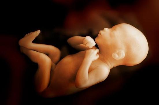
Child's immune system
Its formation begins in the very first weeks of gestation. A baby's immune system continues to develop after birth. The laying of the main protective elements is carried out in the thymus and bone marrow of the fetus. While the baby is in the mother's womb, his body encounters a small number of microorganisms. In this regard, its defense mechanisms are inactive. Before birth, the baby is protected from infections by the mother's immunoglobulins. If any factors adversely affect it, then the correct formation and development of the baby’s defenses may be disrupted. After birth, in this case, the child may get sick more often than other children. But things could happen differently. For example, during pregnancy, the mother of the child can suffer from an infectious disease. And the fetus can develop a strong immunity to this pathology.
After birth, the body is attacked by a huge number of microbes. The immune system must resist them. During the first years of life, the body's protective structures undergo a kind of “training” to recognize and destroy antigens. At the same time, contacts with microorganisms are remembered. As a result, “immunological memory” is formed. It is necessary for a faster manifestation of the reaction to already known antigens. It must be assumed that a newborn’s immunity is weak and he is not always able to cope with danger. In this case, antibodies received in utero from the mother come to the rescue. They are present in the body for approximately the first four months of life. Over the next two months, the proteins received from the mother are gradually destroyed. Between four and six months, the baby is most susceptible to illness. Intensive formation of a child’s immune system occurs before the age of seven. During development, the body becomes acquainted with new antigens. During this entire period, the immune system is trained and prepared for adult life.
How to help a fragile body?
Experts recommend taking care of your child’s immune system even before birth. This means that the expectant mother needs to strengthen her protective structure. During the prenatal period, a woman needs to eat properly, take special microelements and vitamins. Moderate physical activity is also important for immunity. In the first year of life, a child needs to receive breast milk. It is recommended to continue breastfeeding until at least 4-5 months. With milk, protective elements penetrate into the baby’s body. During this period they are very important for immunity. You can even put milk in your child’s nose during a flu epidemic. It contains a lot of useful compounds and will help the baby cope with negative factors.
Additional Methods
Training the immune system can be done in various ways. The most common are hardening, massage, gymnastics in a well-ventilated area, sun and air baths, and swimming. There are also various remedies for immunity. One of them is vaccinations. They have the ability to activate protective mechanisms and stimulate the production of immunoglobulins. Thanks to the introduction of special serums, a memory of the body’s structures to the injected material is formed. Another means for immunity are special medications. They stimulate the activity of the body's protective structure. These medications are called immunostimulants. These are interferon preparations (Laferon, Reaferon), interferonogens (Poludan, Abrizol, Prodigiozan), leukopoiesis stimulators - Methyluracil, Pentoxyl, immunostimulants of microbial origin - Prodignozan, Pyrogenal. , “Bronchomunal”, immunostimulants of plant origin - Schisandra tincture, Eleutherococcus extract, vitamins and much more. etc.
Only an immunologist or pediatrician can prescribe these medications. Independent use of drugs in this group is highly discouraged.
Faculty Control
Department "Humanitarian and social disciplines"
by discipline Physical culture
"The body's immune system
person"
Completed by: student Shundakova K.M.
Group ED20.1/B-12
Checked by Orlov A.N.
Moscow, 2013
The immune system is a collection of organs, tissues and cells, the work of which is aimed directly at protecting the body from various diseases and at destroying foreign substances that have already entered the body.
This system is an obstacle to infections (bacterial, viral, fungal). When the immune system malfunctions, the likelihood of developing infections increases, which also leads to the development of autoimmune diseases, including multiple sclerosis.
Organs included in the human immune system: lymph glands (nodes), tonsils, thymus gland (thymus), bone marrow, spleen and lymphoid formations of the intestine (Peyer's patches). The main role is played by a complex circulation system, which consists of lymphatic ducts connecting the lymph nodes.
A lymph node is a soft tissue formation, oval in shape and 0.2 - 1.0 cm in size, which contains a large number of lymphocytes.
Tonsils are small collections of lymphoid tissue located on both sides of the pharynx. The spleen is very similar in appearance to a large lymph node. The functions of the spleen are varied, it is a filter for blood, a storage for blood cells, and the production of lymphocytes. It is in the spleen that old and defective blood cells are destroyed.
The thymus gland (thymus) is located behind the breastbone. Lymphoid cells in the thymus multiply and “learn.” In children and young people, the thymus is active; the older a person is, the less active the thymus becomes and decreases in size.
Bone marrow is soft, spongy tissue located inside tubular and flat bones. The main task of the bone marrow is the production of blood cells: leukocytes, erythrocytes, platelets.
Peyer's patches - This is a concentration of lymphoid tissue in the intestinal wall. The main role is played by the circulation system, consisting of lymphatic ducts that connect the lymph nodes and transport lymphatic fluid.
Lymphatic fluid (lymph) is a colorless liquid that flows through the lymphatic vessels; it contains many lymphocytes - white blood cells involved in protecting the body from disease.
Lymphocytes are figuratively speaking “soldiers” of the immune system; they are responsible for the destruction of foreign organisms or diseased cells (infected, tumor, etc.). The most important types of lymphocytes (B lymphocytes and T lymphocytes) work together with other immune cells and prevent foreign substances (infections, foreign proteins, etc.) from invading the body. At the first stage, the body “teaches” T-lymphocytes to distinguish foreign proteins from normal (its own) proteins of the body. This learning process takes place in the thymus gland during childhood, since at this age the thymus gland is most active. Then a person reaches adolescence, and the thymus decreases in size and loses its activity.
The immune system appeared along with multicellular organisms and evolved as an aid to their survival. It connects organs and tissues that guarantee the body’s protection from genetically foreign cells and substances that come from the environment. In terms of organization and functioning mechanisms, it is similar to the nervous system.
Both systems are represented by central and peripheral organs that are capable of responding to different signals, have a large number of receptor structures, and specific memory.
The central organs of the immune system include the red bone marrow, and the peripheral organs include the lymph nodes, spleen, tonsils, and appendix.
The central place among the cells of the immune system is occupied by various lymphocytes. When in contact with foreign bodies, with their help, the immune system is able to provide different forms of immune response: the formation of specific blood antibodies, the formation of different types of lymphocytes.
The very concept of immunity was introduced into modern science by the Russian scientist I.I. Mechnikov and the German - P. Ehrlich, who studied the body's defense reactions in the fight against various diseases, primarily infectious ones. Their joint work in this area was even awarded the Nobel Prize in 1908. The work of the French scientist Louis Pasteur, who developed a vaccination method against a number of dangerous infections, also made a great contribution to the science of immunology.
The word immunity comes from the Latin immunis, which means “free from anything.” At first it was believed that immunity protects the body only from infectious diseases. However, research by the English scientist P. Medawar in the mid-twentieth century proved that immunity provides protection in general from any foreign and harmful interference in the human body.
Currently, immunity is understood, firstly, as the body’s resistance to infections, and, secondly, as the body’s responses aimed at destroying and removing from it everything that is alien to it and poses a threat. It is clear that if people did not have immunity, they simply would not be able to exist, and its presence allows us to successfully fight diseases and live to old age.
The immune system has been formed over many years of human evolution and acts like a well-oiled mechanism and helps fight diseases and harmful environmental influences. Its tasks include recognizing, destroying and removing from the body both foreign agents penetrating from the outside, as well as decay products formed in the body itself (during infectious and inflammatory processes), as well as pathologically changed cells.
The immune system is able to recognize many “strangers”. Among them are viruses, bacteria, toxic substances of plant or animal origin, protozoa, fungi, and allergens. She includes among them the cells of one’s own body that have turned cancerous and therefore become “enemies.” Its main goal is to provide protection from all these “strangers” and preserve the integrity of the internal environment of the body, its biological individuality.
How is “enemies” recognized? This process occurs at the genetic level. The fact is that each cell carries its own genetic information, unique only to a given person (we can call it a mark). This is what the immune system analyzes when it detects penetration into the body or changes in it. If the information matches (the label is present), then it is yours; if it does not match (the label is missing), it means it is someone else’s.
In immunology, foreign agents are usually called antigens. When the immune system detects them, defense mechanisms immediately turn on, and the fight against the “stranger” begins. Moreover, to destroy each specific antigen, the body produces specific cells, they are called antibodies. They fit antigens like a key to a lock. Antibodies bind to the antigen and eliminate it - this is how the body fights the disease.
One of the immune reactions is allergy - a state of increased response of the body to allergens. Allergens are substances or objects that contribute to the occurrence of an allergic reaction in the body. They are divided into internal and external.
External allergens include some foods (eggs, chocolate, citrus fruits), various chemicals (perfumes, deodorants), and medications.
Internal allergens are the body’s own tissues, usually with altered properties. For example, with burns, the body perceives dead tissue as foreign and creates antibodies for them. The same reactions can occur with the bites of bees, bumblebees, and other insects. Allergic reactions develop rapidly or sequentially. When an allergen affects the body for the first time, antibodies with increased sensitivity to it are produced and accumulated. When this allergen enters the body again, an allergic reaction occurs, for example, skin rashes and various tumors appear.


