Antiphospholipid syndrome - symptoms and treatment methods. Treatment of antiphospholipid syndrome modern recommendations Antiphospholipid syndrome treatment and prevention
Antiphospholipid syndrome (APS) is a clinical and laboratory symptom complex that includes venous and/or arterial thrombosis, various forms of obstetric pathology (primarily recurrent miscarriage), thrombocytopenia, as well as various other neurological, skin, cardiovascular and hematological disorders. A characteristic immunological feature of APS is antibodies to phospholipids - a heterogeneous group of antibodies that react with a wide range of phospholipids and phospholipid-binding proteins. APS most often develops in SLE (secondary APS) or in the absence of another leading disease (primary APS).
The true prevalence of APS in the population is still unknown. The frequency of detection of antibodies to phospholipids in the serum of healthy people varies from 0 to 14%, on average 2-4% (in high titers in less than 0.2%). The disease often develops at a young age and can occur in children and even newborns. In elderly people, the development of APS may be associated with malignant neoplasms. In the general population, APS is more often detected in women. However, among patients with primary APS, an increase in the proportion of men is noted.
ETIOLOGY
The causes of APS are not known. An increase in the level (usually transient) of antibodies to phospholipids is observed against the background of a wide range of bacterial and viral infections. However, thrombotic complications in patients with infections develop less frequently than antibodies to phospholipids are detected. There is evidence of an immunogenetic predisposition to overproduction of antibodies to phospholipids. There was an increase in the frequency of detection of antibodies to phospholipids in families of patients with APS; Cases of APS (usually primary) have been described in members of the same family.
PATHOGENESIS
Abs to phospholipids bind to phospholipids in the presence of a cofactor, which is β 2 -glycoprotein I, a protein that binds to phospholipids and has anticoagulant activity. Antiphospholipid antibodies present in the serum of patients with APS react with Ags formed during the interaction of phospholipid components of the membranes of endothelial and other cells (platelets, neutrophils) and β 2 -glycoprotein I. As a result of this interaction, the synthesis of anticoagulants (prostacyclin, antithrombin III) is suppressed , annexin V, etc.) and increased formation of procoagulant (thromboxane, tissue factor, platelet activating factor, etc.) mediators, activation of the endothelium (expression of adhesion molecules) and platelets is induced, and neutrophil degranulation occurs.
Antiphospholipid antibodies detected in the serum of patients with infectious diseases usually react with phospholipids in the absence of β 2 -glycoprotein I and do not have the properties described above.
CLASSIFICATION
The following clinical and laboratory variants of APS are distinguished.
Primary APS.
Secondary APS.
. "Catastrophic" APS.
In some patients, APS manifests itself predominantly as venous thrombosis, in others as a stroke, in others as obstetric pathology or thrombocytopenia. The development of APS does not correlate with the activity of the underlying disease. Approximately half of patients with APS suffer from the primary form of the disease. However, the question of the nosological independence of primary APS is not completely clear. Primary APS can sometimes be a variant of the onset of SLE. On the contrary, in some patients with classical SLE at the onset, signs of APS may subsequently come to the fore.
In some patients, APS may present with acute recurrent coagulopathy and vasculopathy affecting vital organs and resembling disseminated intravascular coagulation or hemolytic uremic syndrome. This condition is called "catastrophic" APS.
CLINICAL PICTURE
Since APS is based on non-inflammatory thrombotic damage to vessels of any size and location, the range of clinical manifestations is extremely diverse.
Venous thrombosis is the most common manifestation of APS. Thrombi are usually localized in the deep veins of the lower extremities, but are often found in the hepatic, portal veins, superficial veins, etc. Repeated pulmonary embolisms from the deep veins of the lower extremities are typical, sometimes leading to pulmonary hypertension. APS (more often primary than secondary) is the second most common cause of Budd-Chiari syndrome. Thrombosis of the central vein of the adrenal glands can lead to adrenal insufficiency.
Arterial thrombosis. Thrombosis of intracerebral arteries, leading to stroke and transient ischemic attacks, is the most common localization of arterial thrombosis in APS. Recurrent ischemic strokes caused by damage to small vessels sometimes occur without significant neurological disorders and can manifest as convulsive syndrome, multi-infarct dementia (resembling Alzheimer's disease), and mental disorders.
A variant of APS is Sneddon syndrome, manifested by recurrent thrombosis of cerebral vessels, livedo reticularis, hypertension and developing in young and middle-aged people. Other neurological disorders include migraine headaches, epileptiform seizures, chorea, and transverse myelitis. Sometimes neurological disorders in APS resemble those in multiple sclerosis.
Heart valve damage is one of the common cardiac manifestations of APS. It varies from minimal disturbances detected only by echocardiography (slight regurgitation, thickening of the valve leaflets) to severe heart defects (stenosis or insufficiency of the mitral, less often the aortic and tricuspid valves). Some patients quickly develop severe valve damage with vegetations caused by thrombotic overlays, similar to valve damage in infective endocarditis. Detection of vegetations on the valves, especially if they are combined with hemorrhages in the nail bed and fingers in the form of “drumsticks”, dictates the need for differential diagnosis with infective endocarditis. The development of blood clots in the cardiac cavity, simulating cardiac myxoma, has been described. One of the possible localizations of arterial thrombosis associated with the synthesis of antibodies to phospholipids is the coronary arteries (in men with SLE this is the most common localization).
Frequent complications of APS include hypertension. It can be labile and is often associated with livedo reticularis and damage to the cerebral arteries as part of Sneddon syndrome, or stable, malignant, manifested by symptoms of hypertensive encephalopathy. The development of hypertension in APS can be associated with many causes, including renal vascular thrombosis, renal infarction, thrombosis of the abdominal aorta (pseudocoarctation), and intraglomerular thrombosis. A connection has been noted between the overproduction of antibodies to phospholipids and the development of fibromuscular dysplasia of the renal arteries. A rare complication of APS is thrombotic pulmonary hypertension, which is associated with both recurrent pulmonary embolism and local ( in situ) thrombosis of the pulmonary vessels.
Kidney damage in APS is associated with intraglomerular microthrombosis and is referred to as renal thrombotic microangiopathy. Microthrombosis of the glomeruli of the kidneys is considered the cause of the subsequent development of glomerulosclerosis, leading to dysfunction of the organ.
Obstetric pathology is considered one of the most characteristic signs of APS: recurrent miscarriage, recurrent spontaneous abortions, intrauterine fetal death, preeclampsia. Fetal loss can occur at any stage of pregnancy, but more often in the second and third trimesters.
Skin lesions in APS are characterized by a variety of clinical manifestations (usually livedo reticularis). Less common are skin ulcers and pseudovasculitic lesions (purpura, palmar and plantar erythema, pustules, gangrene of the fingers).
Thrombocytopenia is a typical hematological sign of APS. The development of hemorrhagic complications is rare and, as a rule, is associated with a concomitant defect in blood coagulation factors, kidney pathology, or an overdose of anticoagulants. Hemolytic anemia with a positive Coombs test is often observed; Evans syndrome (a combination of thrombocytopenia and hemolytic anemia) is less common.
LABORATORY RESEARCH
Laboratory diagnosis of APS is based on the determination of lupus anticoagulant using functional tests and antibodies to cardiolipin using ELISA. In general, lupus anticoagulant has higher specificity, and anticardiolipin antibodies have higher sensitivity for diagnosing APS. Lupus anticoagulant and antibodies to cardiolipin are detected in 30-40% and 40-50% of patients with SLE, respectively. In the presence of antibodies to phospholipids, the risk of developing thrombosis is 40%, while in the absence of antibodies it is no higher than 15%. A method has been developed for determining antibodies that react with β 2 -glycoprotein I, an increase in the level of which correlates better with the development of thrombosis than an increase in the level of antibodies to phospholipids. The course of APS, the severity and prevalence of thrombotic complications in most cases do not depend on the concentration of antibodies to phospholipids.
DIAGNOSTICS
For diagnosis, preliminary criteria for the classification of APS should be used (Table 46-1).
Table 46-1. Criteria for diagnosing APS
. Clinical criteria | . Laboratory criteria |
Vascular thrombosis 1 or more episodes of thrombosis of blood vessels supplying any organ or tissue. With the exception of superficial vein thrombosis, thrombosis must be confirmed by angiography, ultrasound or morphological method. With morphological confirmation, signs of thrombosis should be observed in the absence of pronounced inflammatory infiltration of the vascular wall. | Anti-cardiolipin antibodies of the IgG or IgM class in medium or high titers, determined at least 2 times within 6 weeks using an ELISA that allows the determination of antibodies to β 2 -glycoprotein |
Obstetric pathology 1 or more unexplained deaths of a morphologically normal fetus before the 10th month of gestation 1 or more deaths of a morphologically normal fetus before the 34th week of gestation due to severe preeclampsia or eclampsia or severe placental insufficiency 3 or more unexplained consecutive spontaneous abortions before the 10th week of gestation, with the exception of anatomical and hormonal disorders of the maternal reproductive system or chromosomal abnormalities in the mother or father | Lupus anticoagulant detected at least 2 times within 6 weeks using a standardized method including the following steps Prolongation of phospholipid-dependent blood coagulation using screening tests (APTT, kaolin test, Russell's viper test, prothrombin time, textarine time) When mixed with normal plasma without platelets, the prolongation of blood clotting time according to screening tests is maintained. Normalization of blood clotting time by adding excess phospholipids Rule out other coagulopathies (factor VIII inhibitors or heparin) |
To make a reliable diagnosis of APS, a combination of at least one clinical and one laboratory criterion is necessary.
APS should be suspected in cases of thrombotic disorders (especially multiple, recurrent, with unusual localization), thrombocytopenia, obstetric pathology in young and middle-aged people, as well as in unexplained thrombosis in newborns, in case of skin necrosis during treatment with indirect anticoagulants and in patients with prolonged aPTT during a screening study. With APS, a large number of pseudo-syndromes are observed, which can imitate vasculitis, infective endocarditis, heart tumors, multiple sclerosis, hepatitis, nephritis, etc.
TREATMENT
Prevention and treatment of APS is a complex task (Table 46-2). This is due to the heterogeneity of the pathogenetic mechanisms underlying APS, the polymorphism of clinical manifestations, and the lack of reliable clinical and laboratory indicators to predict the recurrence of thrombotic disorders. The risk of recurrent thrombosis is especially high in young patients with persistent high levels of antibodies to cardiolipin, lupus anticoagulant and with simultaneous detection of antibodies to cardiolipin and lupus anticoagulant, as well as in the presence of recurrent thrombosis and/or obstetric pathology in the anamnesis, in the presence of other thrombotic risk factors disorders (hypertension, hyperlipidemia, smoking, taking oral contraceptives), with high activity of SLE, with rapid withdrawal of indirect anticoagulants, with a combination of high titers of antibodies to phospholipids with other coagulation disorders.
. Groups patients | |
Without clinical signs of APS, but with a high level of antibodies to phospholipids | Without risk factors - low doses of acetylsalicylic acid (less than 100 mg/day) ± hydroxychloroquine (100-200 mg/day) (for secondary APS) |
If there are risk factors - warfarin (INR less than 2) ± hydroxychloroquine (100-200 mg/day) |
|
With venous thrombosis | Warfarin (INR=2-3) ± hydroxychloroquine (100-200 mg/day) |
With arterial thrombosis | Warfarin (INR more than 3) ± hydroxychloroquine ± acetylsalicylic acid in low doses (depending on the risk of recurrent thrombosis or bleeding) |
With repeated thromboses | Warfarin (INR greater than 3) ± hydroxychloroquine ± low-dose acetylsalicylic acid |
In addition, there are a number of features in the treatment of APS.
In patients with high levels of antibodies to phospholipids in the serum, but without clinical signs of APS (including pregnant women without a history of obstetric pathology), they are limited to prescribing small doses of acetylsalicylic acid (75 mg/day). These individuals require careful follow-up, since their risk of thrombotic complications is very high. Moderate thrombocytopenia usually does not require treatment or is controlled with small doses of GCs.
Management of patients with definite APS is based on the prescription of vitamin K antagonists (warfarin) and antiplatelet agents (low doses of acetylsalicylic acid), which are widely used for the prevention of thrombosis not associated with APS. In patients with both secondary and primary APS, treatment with warfarin, which maintains the INR at a level of 2-3 (or more), leads to a significant reduction in the incidence of recurrent thrombotic complications. However, the use of warfarin is associated with a high risk of bleeding. It is advisable to prescribe antimalarial drugs, which, along with an anti-inflammatory effect, have antithrombotic (suppress platelet aggregation and adhesion, reduce the size of a blood clot) and lipid-lowering activity.
The use of warfarin during pregnancy is contraindicated, as this leads to the development of warfarin embryopathy, characterized by impaired growth of the epiphyses of the bones and hypoplasia of the nasal septum, as well as neurological disorders. Treatment with heparin (especially low molecular weight heparins in standard doses) in combination with low doses of acetylsalicylic acid in women with recurrent miscarriage can increase the frequency of successful births by approximately 2-3 times and is significantly more effective than glucocorticoid therapy.
Treatment with GCs and cytotoxic drugs is not recommended, except in cases of “catastrophic” APS.
FORECAST
The development of APS in SLE reduces patient survival. Prognostically unfavorable factors for recurrent thrombosis include thrombocytopenia, arterial thrombosis, and a persistent increase in the level of antibodies to cardiolipin.
All cells of the body contain esters of higher fatty acids and polyhydric alcohols. These chemical compounds are called phospholipids; they are responsible for maintaining the proper structure of tissues, participate in metabolic processes and the breakdown of cholesterol. The general state of health depends on the concentration of these substances.
APS syndrome - what is it?
About 35 years ago, rheumatologist Graham Hughes discovered a pathology in which the immune system begins to produce specific antibodies against phospholipids. They attach to platelets and vascular walls, interact with proteins, and enter into metabolic and blood clotting reactions. Both secondary and primary antiphospholipid antibody syndrome are autoimmune diseases of unknown origin. Young women of reproductive age are more susceptible to this problem.
Antiphospholipid syndrome - causes
Rheumatologists have not yet been able to establish why the disease in question occurs. There is information that antiphospholipid syndrome is more often diagnosed in relatives with a similar disorder. In addition to heredity, experts suggest several other factors that provoke pathology. In such cases, secondary APS develops - the reasons for the production of antibodies are the progression of other diseases that affect the functioning of the immune system. The treatment strategy depends on the mechanisms of the disease.
Primary antiphospholipid syndrome
This type of pathology develops independently, and not against the background of any disorders in the body. This antiphospholipid antibody syndrome is difficult to treat due to the lack of precipitating factors. Often the primary form of the disease is almost asymptomatic and is diagnosed already in the later stages of progression or when complications occur.
This type of autoimmune reaction develops due to the presence of other systemic diseases or certain clinical events. Even conception can be the trigger for the onset of pathological antibody production. Antiphospholipid syndrome in pregnant women occurs in 5% of cases. If the disease in question was diagnosed earlier, pregnancy will significantly worsen its course.

Diseases that are thought to cause antiphospholipid syndrome:
- viral and bacterial infections;
- oncological neoplasms;
- periarteritis nodosa;
- systemic lupus erythematosus.
Antiphospholipid syndrome - symptoms in women
The clinical picture of the pathology is very diverse and nonspecific, which makes differential diagnosis difficult. Sometimes the disorder occurs without any symptoms, but more often antiphospholipid syndrome manifests itself in the form of recurrent thrombosis of superficial and deep blood vessels (arteries or veins):
- lower extremities;
- liver;
- retina;
- brain;
- hearts;
- lungs;
- kidney
Common symptoms in women:
- pronounced vascular pattern on the skin (livedo reticularis);
- myocardial infarction;
- migraines;
- suffocation;
- chest pain;
- varicose veins;
- thrombophlebitis;
- stroke;
- arterial hypertension;
- acute renal failure;
- ascites;
- severe dry cough;
- necrosis of bones and soft tissues;
- portal hypertension;
- gastrointestinal bleeding;
- severe liver damage;
- splenic infarction;
- intrauterine fetal death;
- spontaneous miscarriage.
Antiphospholipid syndrome - diagnosis
It is difficult to confirm the presence of the described pathology, because it disguises itself as other diseases and has nonspecific symptoms. To diagnose the disease, doctors use 2 groups of classification criteria. Testing for antiphospholipid syndrome first involves taking a medical history. The first type of assessment indicators includes clinical phenomena:
- Vascular thrombosis. The medical history must contain one or more cases of damage to veins or arteries, established instrumentally and laboratory.
- Obstetric pathology. The criterion is taken into account if intrauterine fetal death occurred after the 10th week of gestation or was observed until the 34th week of pregnancy in the absence of chromosomal, hormonal and anatomical defects on the part of the parents.
After collecting anamnesis, the doctor prescribes additional tests. Antiphospholipid syndrome is confirmed when a combination of one clinical symptom and a laboratory criterion (minimum) is present. In parallel, a number of differential diagnostic measures are carried out. To do this, the specialist recommends undergoing examinations to rule out similar diseases.

Antiphospholipid syndrome - analysis
The study of biological fluids helps to identify laboratory signs of this disorder. The doctor prescribes a blood test for antiphospholipid syndrome to determine the presence of antibodies to cardiolipins and lupus anticoagulant in the plasma and serum. Additionally, the following may be found:
- cryoglobulins;
- antibodies to red blood cells;
- T- and B-lymphocytes in increased concentration;
- antinuclear and rheumatoid factor.
- HLA-DR7;
- HLA-B8;
- HLA-DR2;
- DR3-HLA.
How to treat antiphospholipid syndrome?
Treatment for this autoimmune disorder depends on its form (primary, secondary) and the severity of clinical signs. Difficulties arise if a pregnant woman is diagnosed with antiphospholipid syndrome - treatment should effectively relieve the symptoms of the disease, prevent thrombosis, and at the same time not pose a danger to the fetus. To achieve lasting improvements, rheumatologists use a combination therapeutic approach.
Can antiphospholipid syndrome be cured?
It is impossible to completely get rid of the described problem until the causes of its occurrence have been established. For antiphospholipid syndrome, it is necessary to use complex treatment aimed at reducing the amount of corresponding antibodies in the blood and preventing thromboembolic complications. In severe cases of the disease, anti-inflammatory therapy is required.
The main way to eliminate the signs of this pathology is the use of antiplatelet agents and indirect anticoagulants:
- acetylsalicylic acid (Aspirin and analogues);
- Acenocoumarol;
- Phenilin;
- Dipyridamole.
How to treat antiphospholipid syndrome - clinical recommendations:
- Stop smoking, drinking alcohol and drugs, and oral contraceptives.
- Adjust your diet in favor of foods rich in vitamin K - green tea, liver, leafy green vegetables.
- Get plenty of rest and follow a daily routine.

If standard therapy is ineffective, additional medications are prescribed:
- aminoquinolines – Plaquenil, Delagil;
- direct anticoagulants - Fraxiparine;
- glucocorticoids – , Methylprednisolone;
- platelet receptor inhibitors – Tagren, Clopidogrel;
- heparinoids - Emeran, Sulodexide;
- – Endoxan, Cytoxan;
- immunoglobulins (intravenous administration).
Traditional medicine for antiphospholipid syndrome
There are no effective alternative treatments; the only option is to replace acetylsalicylic acid with natural raw materials. Antiphospholipid syndrome cannot be treated with folk remedies, because natural anticoagulants have too mild an effect. It is important to consult a rheumatologist before using any alternative remedies. Only a specialist will help alleviate antiphospholipid syndrome - the doctor’s recommendations should be strictly followed.
Tea with aspirin properties
Ingredients:- dry white willow bark - 1-2 teaspoons;
- boiling water – 180-220 ml.
Preparation, use:
- Rinse the plant material thoroughly and chop it.
- Brew willow bark with boiling water and leave for 20-25 minutes.
- Drink the solution as tea 3-4 times a day, you can sweeten it to taste.
Antiphospholipid syndrome - prognosis
All rheumatologist patients with the presented diagnosis should be observed for a long time and undergo regular preventive examinations. How long you can live with antiphospholipid syndrome depends on its form, severity and the presence of concomitant immunological disorders. If primary APS with moderate symptoms is detected, timely therapy and preventive treatment help to avoid complications, the prognosis in such cases is as favorable as possible.
Aggravating factors are the combination of the disease in question with lupus erythematosus, thrombocytopenia, persistent arterial hypertension and other pathologies. In these situations, antiphospholipid complex syndrome (catastrophic) often develops, which is characterized by an increase in clinical signs and recurrent thrombosis. Some consequences can be fatal.
Antiphospholipid syndrome and pregnancy
The described disease is a common cause of miscarriage, so all expectant mothers should undergo a preventive examination and donate blood for a coagulogram. Antiphospholipid syndrome in obstetrics is considered a serious factor provoking fetal death and miscarriage, but its presence is not a death sentence. A woman with this diagnosis is able to bear and give birth to a healthy baby if during pregnancy she follows all the doctor’s recommendations and takes antiplatelet drugs.
A similar scheme is used when artificial insemination is planned. Antiphospholipid syndrome and IVF are quite compatible, you just have to first take a course of antithrombotic drugs. The use of anticoagulants and antiplatelet agents will continue throughout the gestation period. The effectiveness of this treatment is close to 100%.
Autoimmune pathology, which is based on the formation of antibodies to phospholipids, which are the main lipid components of cell membranes. Antiphospholipid syndrome can manifest itself as venous and arterial thrombosis, arterial hypertension, valvular heart defects, obstetric pathology (recurrent miscarriage, intrauterine fetal death, preeclampsia), skin lesions, thrombocytopenia, hemolytic anemia. The main diagnostic markers of antiphospholipid syndrome are antibodies to cardiolipin and lupus anticoagulant. Treatment of antiphospholipid syndrome comes down to the prevention of thrombosis, the prescription of anticoagulants and antiplatelet agents.

General information
Antiphospholipid syndrome (APS) is a complex of disorders caused by an autoimmune reaction to phospholipid structures present on cell membranes. The disease was described in detail by the English rheumatologist Hughes in 1986. There are no data on the true prevalence of antiphospholipid syndrome; It is known that insignificant levels of antibodies to phospholipids in blood serum are found in 2-4% of practically healthy individuals, and high titers - in 0.2%. Antiphospholipid syndrome is diagnosed 5 times more often among young women (20-40 years), although men and children (including newborns) can suffer from the disease. As a multidisciplinary problem, antiphospholipid syndrome (APS) attracts the attention of specialists in the field of rheumatology, obstetrics and gynecology, and cardiology.
Reasons
The underlying causes of antiphospholipid syndrome are unknown. Meanwhile, factors predisposing to increased levels of antibodies to phospholipids have been studied and identified. Thus, a transient increase in antiphospholipid antibodies is observed against the background of viral and bacterial infections (hepatitis C, HIV, infectious mononucleosis, malaria, infective endocarditis, etc.). High titers of antibodies to phospholipids are found in patients with systemic lupus erythematosus, rheumatoid arthritis, Sjögren's disease, periarteritis nodosa, and autoimmune thrombocytopenic purpura.
Overproduction of antiphospholipid antibodies can occur with malignant neoplasms, taking medications (psychotropic drugs, hormonal contraceptives, etc.), and discontinuation of anticoagulants. There is information about a genetic predisposition to increased synthesis of antibodies to phospholipids in individuals who carry the HLA DR4, DR7, DRw53 antigens and in relatives of patients with antiphospholipid syndrome. In general, the immunobiological mechanisms of the development of antiphospholipid syndrome require further study and clarification.
Depending on the structure and immunogenicity, “neutral” (phosphatidylcholine, phosphatidylethanolamine) and “negatively charged” (cardiolipin, phosphatidylserine, phosphatidylinositol) phospholipids are distinguished. The class of antiphospholipid antibodies that react with phospholipids includes lupus anticoagulant, antibodies to cardiolipin, beta2-glycoprotein-1-cofactor-dependent antiphospholipids, etc. By interacting with phospholipids of the membranes of vascular endothelial cells, platelets, neutrophils, antibodies cause hemostasis disturbances, expressed in the tendency to hypercoagulation.
Classification
Taking into account the etiopathogenesis and course, the following clinical and laboratory variants of antiphospholipid syndrome are distinguished:
- primary– there is no connection with any underlying disease that can induce the formation of antiphospholipid antibodies;
- secondary- antiphospholipid syndrome develops against the background of another autoimmune pathology;
- catastrophic– acute coagulopathy, occurring with multiple thromboses of internal organs;
- AFL-negative a variant of antiphospholipid syndrome, in which serological markers of the disease (Abs to cardiolipin and lupus anticoagulant) are not detected.
Symptoms of antiphospholipid syndrome
According to modern views, antiphospholipid syndrome is an autoimmune thrombotic vasculopathy. In APS, damage can affect vessels of various sizes and locations (capillaries, large venous and arterial trunks), which causes an extremely diverse range of clinical manifestations, including venous and arterial thrombosis, obstetric pathology, neurological, cardiovascular, skin disorders, thrombocytopenia.
The most common and typical sign of antiphospholipid syndrome is recurrent venous thrombosis: thrombosis of the superficial and deep veins of the lower extremities, hepatic veins, portal vein of the liver, retinal veins. Patients with antiphospholipid syndrome may experience recurrent episodes of pulmonary embolism, pulmonary hypertension, superior vena cava syndrome, Budd-Chiari syndrome, and adrenal insufficiency. Venous thrombosis in antiphospholipid syndrome develops 2 times more often than arterial thrombosis. Among the latter, thrombosis of the cerebral arteries predominates, leading to transient ischemic attacks and ischemic stroke. Other neurological disorders may include migraine, hyperkinesis, seizure syndrome, sensorineural hearing loss, ischemic optic neuropathy, transverse myelitis, dementia, and mental disorders.
Damage to the cardiovascular system in antiphospholipid syndrome is accompanied by the development of myocardial infarction, intracardiac thrombosis, ischemic cardiomyopathy, and arterial hypertension. Quite often, damage to the heart valves is observed - from minor regurgitation, detected by echocardiography, to mitral, aortic, tricuspid stenosis or insufficiency. As part of the diagnosis of antiphospholipid syndrome with cardiac manifestations, differential diagnosis with infective endocarditis and cardiac myxoma is required.
Renal manifestations may include both mild proteinuria and acute renal failure. On the part of the gastrointestinal tract, antiphospholipid syndrome causes hepatomegaly, gastrointestinal bleeding, occlusion of mesenteric vessels, portal hypertension, and splenic infarction. Typical lesions of the skin and soft tissues are represented by livedo reticularis, palmar and plantar erythema, trophic ulcers, gangrene of the fingers; musculoskeletal system - aseptic necrosis of bones (femoral head). Hematological signs of antiphospholipid syndrome are thrombocytopenia, hemolytic anemia, and hemorrhagic complications.
In women, APS is often detected in connection with obstetric pathology: repeated spontaneous abortion at various times, intrauterine growth retardation, placental insufficiency, gestosis, chronic fetal hypoxia, premature birth. When managing pregnancy in women with antiphospholipid syndrome, the obstetrician-gynecologist must take into account all possible risks.
Diagnostics
Antiphospholipid syndrome is diagnosed based on clinical (vascular thrombosis, complicated obstetric history) and laboratory data. The main immunological criteria include the detection of medium or high titers of antibodies to cardiolipin of the IgG/IgM class and lupus anticoagulant in the blood plasma twice within six weeks. The diagnosis is considered reliable when at least one main clinical and laboratory criterion is combined. Additional laboratory signs of antiphospholipid syndrome are false-positive RW, positive Coombs test, increased titer of antinuclear factor, rheumatoid factor, cryoglobulins, and antibodies to DNA. A study of CBC, platelets, biochemical blood test, and coagulogram is also indicated.
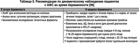
Pregnant women with antiphospholipid syndrome need monitoring of blood coagulation parameters, dynamic ultrasound of the fetus and
Treatment of antiphospholipid syndrome
The main goal of treatment for antiphospholipid syndrome is to prevent thromboembolic complications. Regular moments include moderate physical activity, avoidance of long periods of immobility, participation in traumatic sports and long air travel. Women with antiphospholipid syndrome should not be prescribed oral contraceptives, and should always consult an obstetrician-gynecologist before planning pregnancy. Pregnant patients are advised to take small doses of glucocorticoids and antiplatelet agents, immunoglobulin administration, and heparin injections under the control of hemostasiogram parameters throughout the entire gestation period.
Drug therapy for antiphospholipid syndrome may include the prescription of indirect anticoagulants (warfarin), direct anticoagulants (heparin, nadroparin calcium, enoxaparin sodium), antiplatelet agents (acetylsalicylic acid, dipyridamole, pentoxifylline). Preventive anticoagulant or antiplatelet therapy for most patients with antiphospholipid syndrome is carried out for a long time, and sometimes for life. In the catastrophic form of antiphospholipid syndrome, the administration of high doses of glucocorticoids and anticoagulants, sessions, transfusion of fresh frozen plasma, etc. is indicated.
Forecast
Timely diagnosis and preventive therapy help avoid the development and recurrence of thrombosis, as well as hope for a favorable outcome of pregnancy and childbirth. In case of secondary antiphospholipid syndrome, it is important to monitor the course of the underlying pathology and prevent infections. Unfavorable prognostic factors are the combination of antiphospholipid syndrome with SLE, thrombocytopenia, a rapid increase in the antibody titer to cardiolipin, and persistent arterial hypertension. All patients diagnosed with antiphospholipid syndrome should be under the supervision of a rheumatologist with periodic monitoring of serological markers of the disease and hemostasiogram parameters.
For quotation: Nasonov E.L. Prevention and treatment of antiphospholipid syndrome: current recommendations and prospects // Breast Cancer. 2004. No. 6. P. 377
Institute of Rheumatology, Russian Academy of Medical Sciences, Moscow
Institute of Rheumatology, Russian Academy of Medical Sciences, Moscow
A antiphospholipid syndrome (APS) is a clinical and laboratory symptom complex characterized by venous and arterial thrombosis, pregnancy pathology and some other less common clinical manifestations and laboratory disorders pathogenetically associated with the synthesis of antiphospholipid antibodies (aPL).
Prevention and treatment of APS is a complex and underdeveloped problem. . This is explained by the heterogeneity of the pathogenetic mechanisms underlying APS and the lack of reliable clinical and laboratory indicators that allow predicting the risk of recurrent thrombosis. Currently, there are no generally accepted international standards for the management of patients with various forms of APS, and the proposed recommendations are based mainly on the results of “open” trials or retrospective analyzes of disease outcomes. Approaches to the prevention and treatment of atherosclerotic vascular lesions, which often develop in patients with APS, have not been sufficiently studied. Since “specific” methods for treating the immunopathological disorders underlying APS have not been developed, the management of patients with APS (as with other thrombophilias) is based on the use of anticoagulant (vitamin K antagonists, heparin) and antiplatelet drugs - acetylsalicylic acid (ASA). A characteristic feature of APS is a high risk of recurrent thrombosis . Therefore, most patients are forced to take antiplatelet and/or anticoagulant drugs for a long time, and sometimes for life.
It is believed that the risk of development (and recurrence) of thrombosis in APS can be reduced by eliminating potentially controllable “risk factors”, but the true effectiveness of these recommendations is unknown. Risk factors that must be taken into account when developing patient management tactics are presented in Table 1.
Prevention of thrombosisAcetylsalicylic acid
Considering the definite connection between an increase in aPL titers and the risk of developing thrombosis in the general population, it is believed that a persistent increase in aPL levels (even in the absence of clinical signs of APS) is the basis for the prophylactic administration of low doses of ASA. Data from two retrospective studies evaluating the effectiveness of ASA have recently been published. One study examined 65 women with obstetric pathology associated with APS. During 8 years of follow-up, thrombotic disorders developed in only 3 (10%) of 31 women receiving ASA and in 20 (59%) of 34 women not receiving ASA. Another study of 77 patients with APS or no thrombosis but positive aPL results showed that ASA use was clearly associated with a lower incidence of thrombosis.
Considering the definite connection between an increase in aPL titers and the risk of developing thrombosis in the general population, it is believed that a persistent increase in aPL levels (even in the absence of clinical signs of APS) is the basis for the prophylactic administration of low doses of ASA. Data from two retrospective studies evaluating the effectiveness of ASA have recently been published. One study examined 65 women with obstetric pathology associated with APS. During 8 years of follow-up, thrombotic disorders developed in only 3 (10%) of 31 women receiving ASA and in 20 (59%) of 34 women not receiving ASA. Another study of 77 patients with APS or no thrombosis but positive aPL results showed that ASA use was clearly associated with a lower incidence of thrombosis.Hydroxychloroquine
Aminoquinoline (antimalarial) drugs (hydroxychloroquine) can have a significant preventive effect, at least in secondary APS associated with systemic lupus erythematosus (SLE). Along with the anti-inflammatory, hydroxychloroquine has certain antithrombotic (suppress platelet aggregation and adhesion, reduce the size of the blood clot) and hypolipidemic effects. The use of hydroxychloroquine is clearly indicated for all aPL-positive patients with SLE.
Warfarin
Treatment with vitamin K antagonists (warfarin) is certainly a more effective, but less safe (compared to ASA) method of preventing venous and arterial thrombosis in APS. Let us recall that the use of vitamin K antagonists and anticoagulants requires careful clinical (hemorrhagic complications) and laboratory (determination of prothrombin time) monitoring. To standardize the results of this test, the international normalized ratio (INR) parameter should be assessed, which takes into account the effect of thromboplastin used in the test on the prothrombin time.
The treatment regimen with warfarin for APS is the same as for other thrombophilias, and consists of prescribing a “saturating” dose (5 mg/day) for the first 2 days, and then selecting the optimal dose of the drug, focusing on the “target” INR . It should be remembered that in elderly people, lower doses of warfarin should be used to achieve the same level of anticoagulation than in young people.
Of particular importance is the question of the intensity and duration of anticoagulation. It is known that an increase in INR from 2-3 to 3.1-4.0 is associated with an increase in the frequency of severe hemorrhagic complications (intracranial hemorrhages or hemorrhages leading to death, requiring blood transfusion or hospitalization). Let us remember that to Risk factors for hemorrhagic complications during warfarin treatment include:
- old age (32% increase in the frequency of any bleeding and a 46% increase in the frequency of “major” bleeding every 10 years after 40 years)
- uncontrolled arterial hypertension (systolic blood pressure >180 mmHg, diastolic blood pressure >100 mmHg)
- peptic ulcer
- drinking alcohol
- taking NSAIDs (including low doses of ASA) and paracetamol
- history of stroke
- taking multiple medications
- taking azathioprine
- taking high doses of methylprednisolone
- polymorphism of cytochrome P450СY2C2, responsible for heparin metabolism
- diffuse decrease in the density of the white matter of the brain (detected by MRI or CT).
In the general population of patients with venous thrombosis, discontinuation of warfarin is associated with the same (5-10%) incidence of recurrent thrombosis, regardless of the duration of previous warfarin treatment (6, 12 and 24 months). However, as already noted, APS is characterized by a high risk of recurrent thrombosis. Therefore, patients with APS and venous thrombosis should be treated with warfarin for a longer period (>12 months) than patients without APS (3-6 months).
One group of authors, at the risk of recurrent thrombosis (including ischemic stroke) in patients with APS, recommends intensive anticoagulation with warfarin, which allows maintaining the INR at a level of >3.1. At the same time, other authors indicate the effectiveness (especially in venous thrombosis) of an average level of anticoagulation, which allows maintaining the INR at a level of 2.0-3.0. M.A. Cronther et al. conducted a randomized, double-blind, controlled trial that compared the efficacy and safety of moderate-intensive (INR 2-3) and high-intensity (INR 3.1-4) anticoagulation with warfarin in APS. The study included 114 patients with high/moderate levels of aPL and at least one episode of thrombosis (venous and arterial) in history; The duration of treatment was 2.7 years. During the observation period, recurrent thrombosis occurred in 6 of 56 (10.7%) patients receiving high-intensity therapy and 2 of 58 (3.4%) receiving moderate-intensive warfarin therapy. Interestingly, the incidence of severe bleeding in the compared groups was approximately the same (3 patients who received intensive anticoagulation and 4 who received moderate anticoagulation).
Thus, at present, the most justified use of warfarin in medium doses (INR 2.0-3.0) in patients with the first episode of venous thrombosis in the absence of other risk factors for recurrent thromboembolic complications, while in patients with a history of recurrent thrombosis Intensive anticoagulation (INR >3.0) is probably more justified.
The question of use of warfarin in patients with APS and ischemic stroke . This is due to the fact that, according to numerous controlled studies, warfarin has no advantage over ASA in preventing recurrent stroke in the general population of patients with cerebral strokes and often causes severe intracranial bleeding. However, according to many authors, with APS the risk of repeated cerebral thrombosis is higher than the risk of bleeding. At the same time, the risk of bleeding against the background of intensive anticoagulation in APS can be compensated to a certain extent by the fact that patients with this syndrome are usually young. According to G. Ruiz-Irastorza et al. , in patients with APS during treatment with warfarin, the incidence of “major” bleeding was 6 cases per 100 patient-years, there were no fatal bleedings in any case, and intracranial hemorrhages occurred in only 1 patient. At the same time, relapses of thrombosis developed mainly in patients who had insufficient anticoagulation (INR< 3,0). Таким образом, вопрос об оптимальном уровне антикоагуляции у пациентов с АФС и с ишемическими инсультами остается открытым и должен решаться индивидуально как с учетом тяжести и факторов риска рецидивов тромбоза, так и риска кровотечений .
It should be emphasized that many patients with APS experience spontaneous fluctuations in INR, making it difficult to select an effective and safe dose of warfarin. In this case, fluctuations in INR are associated with taking medications that affect the metabolism of warfarin, many of which are widely used in rheumatology (for example, cytostatics, GCs, allopurinol, NSAIDs, cephalosporins, etc.). In addition, fluctuations in INR may be associated with different properties of thromboplastin, which is used to determine prothrombin time. The dose of indirect anticoagulants is difficult to select in the presence of VA in the blood, the presence of which sometimes leads to “false-positive” results - to an increase in prothrombin time and INR in vitro, in the absence of effective anticoagulation in vivo. Patients with APS often exhibit resistance to warfarin, which is genetic in nature (mutation of coagulation factors V and II).
T.M. Reshetnyak et al. The effectiveness of warfarin was studied in 20 patients (5 men and 15 women) with APS, among whom 8 had primary APS and 12 had APS with SLE. 18 patients received warfarin for a year, and two for 4 years. Patients with a history of arterial thrombosis received pentoxifylline or low doses of ASA (50-100 mg/day).
Patients with APS were divided into three groups. The first group included 8 patients with a target INR of 2.0, the second - 7 with INR of 3.0, and the third - 7 patients with INR of 2.0 who received ASA (100 mg/day) and pentoxifylline (600 to 1200 mg/day. ). Recurrence of venous thrombosis occurred in two patients with INR<2,0. В других группах рецидивов не отмечено. Однако у 2-х пациентов 2 и 3 групп имели место «большие» кровотечения. Частота «малых» геморрагий в сравниваемых группах не различалась.
If warfarin monotherapy is insufficiently effective, combination therapy with indirect anticoagulants and low doses of ASA (and/or dipyridomole) is possible, which is most justified in young people without bleeding risk factors (secondary APS, thrombocytopenia, platelet dysfunction associated with the presence of VA, prothrombin defects ) .
In case of excessive anticoagulation (INR>4.0) in the absence of bleeding, it is recommended to temporarily discontinue warfarin until the INR value returns to the desired level. More rapid normalization of INR can be achieved by administering small doses of vitamin K: 1 mg orally (reduces the risk of at least “minor” bleeding) or 0.5 mg intravenously. High doses of vitamin K should be avoided as this may lead to long-term (several days) resistance to vitamin K antagonists. Subcutaneous injections of vitamin K are not recommended due to the marked variability of absorption. In the case of hypercoagulation, accompanied by “major” bleeding, administration of vitamin K alone is not enough, since the full effect develops only 12-24 hours after administration. In this case, it is recommended to administer fresh frozen plasma or, more preferably, prothrombin complex concentrate.

The central place in the treatment of acute thrombotic complications in APS is occupied by direct anticoagulants - heparin and especially low molecular weight heparin preparations. The tactics of using direct anticoagulants in patients with APS does not differ from the generally accepted ones:
The central place in the treatment of acute thrombotic complications in APS is occupied by direct anticoagulants - heparin and especially low molecular weight heparin preparations. The tactics of using direct anticoagulants in patients with APS does not differ from the generally accepted ones:1. Determine the basal APTT level, prothrombin time and complete blood count.
2. Confirm that there are no contraindications for heparin therapy.
3. Administer 5000 IU of heparin intravenously.
4. Resolve the issue of heparin therapy tactics.
Start a continuous intravenous infusion of unfractionated heparin - 18 IU/kg/hour (on average 30,000/24 hours for a man weighing 70 kg):
Determine aPTT every 6 hours for the first 24 hours, then daily;
Maintain APTT at 1.5-2.5;
Continue infusions for 5-7 days.
Subcutaneous administration of heparin: start with a dose of 17,500 IU every 12 hours (or 250 IU/kg every 12 hours).
5. Determine platelet levels every day due to the possibility of thrombocytopenia.
6. If patients have not previously received warfarin, then it should be prescribed within the first 24-48 hours from the start of heparin therapy.
7. Continue treatment with heparin for at least 4-5 days after prescribing warfarin. In patients with massive ileofemoral thrombosis or pulmonary thromboembolism, treatment with heparin is carried out for at least 10 days.
8. Stop heparin administration when the INR reaches > 2 within 48 hours.
In patients with risk factors for recurrent thrombosis over a long period of time, intensive prophylaxis should be carried out using low molecular weight heparin.
Catastrophic antiphospholipid syndrome The prognosis of catastrophic APS largely depends on how early the diagnosis is made and “aggressive” therapy is initiated. For treatment "catastrophic" APS
The entire arsenal of intensive and anti-inflammatory therapy methods is used to treat critical conditions in rheumatic diseases (Fig. 1).

Rice. 1. Treatment algorithm<катастрофического>AFS
The effectiveness of therapy to a certain extent depends on the ability to eliminate the factors that provoke its development (for example, suppression of infection and/or activity of the underlying disease). If an infection is suspected, antibiotic therapy should be immediately prescribed, and if gangrene of the limbs develops, amputation should be performed. “Nonspecific” intensive therapy is important, for example, hemodialysis in patients with rapidly developing renal failure, ventilation, administration of inotropic drugs, etc.
Carrying out intensive therapy glucocorticoids is not aimed at treating the “thrombotic” disorders themselves, but is determined by the need to manage the “systemic inflammatory response” syndrome. Let us recall that systemic inflammatory response syndrome is characterized by diffuse inflammation of the vascular endothelium associated with overproduction of TNF-a and IL-1. A number of clinical manifestations of APS, associated with both thrombosis of small vessels and widespread necrosis (for example, respiratory distress syndrome in adults, etc.), are indications for the prescription of high doses of glucocorticoids. Typically, standard pulse therapy is recommended (1000 mg methylprednisolone per day for 3-5 days), followed by high doses of glucocorticoids (1-2 mg/kg/day) orally. It should be emphasized once again that glucocorticoids themselves do not affect the risk of developing recurrent thrombosis.
Intravenous immunoglobulin is administered at a dose of 0.4 g/kg for 4-5 days and is especially effective in the presence of thrombocytopenia. It should, however, be remembered that intravenous immunoglobulin can cause renal dysfunction, especially in elderly people who have received nephrotoxic drugs.
“Catastrophic” APS is the only absolute indication for sessions plasmapheresis (it is recommended to remove 2-3 liters of plasma over 3-5 days) in patients with APS, which should be combined with the most intense anticoagulant therapy, the use of fresh frozen plasma for replacement, and, if indicated, with pulse therapy with GC and cyclophosphamide. Plasmapheresis is the method of choice for thrombotic thrombocytopenic purpura and thrombotic microangiopathic hemolytic anemia, which often complicates CAPS.
Cyclophosphamide (0.5-1.0 g per day) is to a certain extent indicated for the development of catastrophic APS against the background of exacerbation of SLE and for the prevention of rebound syndrome after plasmapheresis sessions.
There are no data regarding the possibility of using anticytokines (for example, TNF-a inhibitor). The theoretical basis for their use is data on a significant increase in the level of TNF-a in APS, including catastrophic APS. It is likely that the administration of infliximab could potentially be indicated in a patient with systemic inflammatory response syndrome due to APS.
Pathology of pregnancy The standard for the prevention of recurrent fetal losses (as well as venous and arterial thrombosis in the postpartum period) in APS is the use of low doses of ASA (81 mg/day) in combination with unfractionated heparin or low molecular weight heparin throughout the entire period of pregnancy and for at least 6 months . after childbirth (Table 3).

The main disadvantages of heparin are variable bioavailability when administered subcutaneously and its nonspecific binding to plasma proteins (AT III and coagulation factors), platelet proteins (eg, platelet factor 4) and EC. Moreover, some heparin-binding proteins belong to the proteins of the acute phase of inflammation, the concentration of which increases significantly against the background of inflammation. Finally, another limitation of heparin therapy is a decrease in the ability of heparin to inactivate thrombin, which is complexed with fibrin and factor Xa, associated with activated platelets in the resulting thrombus. Therefore, heparin has no effect on thrombus growth, and after cessation of heparin therapy, a “rebound” increase in coagulation may be observed.
Low molecular weight heparin preparations have advantages over unfractionated heparin in the treatment of venous thrombosis and obstetric pathology in patients with APS and have almost completely replaced the latter (Table 4).

Recently, a randomized trial was conducted that compared the effectiveness of low molecular weight heparin in combination with ASA and intravenous immunoglobulin. The study included 30 women with a history of 3 or more spontaneous abortions. Women receiving heparin and ASA had a higher rate of successful births (84%) than women receiving intravenous immunoglobulin (57%).
During delivery by cesarean section, the administration of low molecular weight heparins is canceled for 2-3 days and resumed in the postpartum period, followed by a transition to taking indirect anticoagulants. Treatment with ASA and heparin reduces the risk of venous and arterial thrombosis, which often develops in patients with APS during and after pregnancy.
It must be borne in mind that long-term heparin therapy in pregnant women can lead to the development of osteoporosis, complicated by skeletal bone fractures. To reduce bone loss, it is recommended to take calcium carbonate (1500 mg) in combination with vitamin D. Treatment with low molecular weight heparin is less likely to lead to osteoporosis than treatment with unfractionated heparin. One of the limitations for the use of low molecular weight heparin is the risk of developing an epidural hematoma during regional anesthesia. Therefore, if preterm labor is expected, treatment with low molecular weight heparin should be discontinued no later than the 36th week of pregnancy.
The use of indirect anticoagulants during pregnancy is in principle contraindicated, as it leads to warfarin embryopathy, characterized by impaired growth of the epiphyses and hypoplasia of the nasal septum, as well as neurological disorders. However, according to a recent study, the administration of warfarin between 15 and 34 weeks of pregnancy in patients with APS (n = 14) was not associated with a teratogenic effect, and the rate of successful birth (86%) was the same as in women taking low doses of ASA and low molecular weight heparin (87%). These data suggest that in some cases, in patients who require active anticoagulant therapy (but cannot tolerate heparin treatment) or have severe systemic thrombosis (stroke, etc.), warfarin may be prescribed from 14 to 34 weeks of pregnancy. In patients undergoing artificial conception or ovulation induction, it is necessary to replace warfarin with heparin. Heparin should be discontinued 12-24 hours before surgery, and therapy should be resumed 6-8 hours later.
Medium/high dose glucocorticoid (GC) treatment, popular in the 1980s, is now largely unused due to side effects in both mother and fetus and lack of evidence of its effectiveness. Moreover, glucocorticoid therapy is associated with severe side effects, including premature membrane rupture, preterm labor, fetal growth restriction, infections, preeclampsia, diabetes, osteopenia, and osteonecrosis. However, before delivery, GCs should not be discontinued in women who received them during pregnancy, and during childbirth they need to additionally administer GCs intravenously in order to avoid adrenal insufficiency. The use of GC is justified in secondary APS (in combination with SLE) and is aimed at treating the underlying disease. Only in some cases, in patients in whom miscarriage cannot be overcome with standard therapy with low doses of ASA and heparin (as well as intravenous immunoglobulin), it is possible to prescribe prednisolone (20-40 mg/day).
The use of intravenous immunoglobulin (0.4 g/kg for 5 days every month) has no advantages over standard treatment with ASA and heparin and is indicated only if “standard” therapy with ASA and heparin is ineffective. There are some preliminary reports of some effectiveness of plasmapheresis, but this method is currently used extremely rarely.
It should be emphasized that the detection of aPL does not affect pregnancy outcomes in women who underwent artificial insemination.
If the presented recommendations are followed, it is possible to increase the frequency of successful births in women with two or more episodes of fetal loss in history to 70-80%. It should, however, be emphasized that even in the case of successful childbirth, patients with APS experience an increase in the incidence of preexlampsia, fetal growth restriction, premature birth and other forms of obstetric pathology. Children of women with APS, as a rule, are born healthy, without signs of impaired physical and neuropsychic development, thrombosis, etc., at least during 5 years of observation.
Hematological disorders Moderate thrombocytopenia, often observed in patients with APS, does not require special treatment. In cases of APS secondary to SLE, thrombocytopenia is usually well controlled with GCs, aminoquinoline drugs, and in resistant cases with low doses of ASA.
Treatment strategy for resistant severe thrombocytopenia (<50000/ мм 3), создающей угрозу кровотечений, до конца не разработана. Этим пациентам, наряду с применением ГК в высоких дозах, целесообразно назначение внутривенного иммуноглобулина. Имеются данные об определенной эффективности препарата даназол (слабый андроген) или дапсон.
In case of ineffectiveness of high doses of GC, the method of “choice” is splenectomy, and in the vast majority of patients stable normalization of platelet levels was noted.
Perioperative management of patients with APS In patients with APS, there is a significant increase in the risk of thrombosis (especially after operations on blood vessels and heart valves) and often the development of catastrophic APS. In general, APS patients constitute a group of very high risk of developing venous thromboembolic complications in the postoperative period.
The development of thrombosis in the pre- and postoperative period may be associated with the following factors:<
- >
- Cancellation of indirect anticoagulants
- Spontaneous increase in coagulability despite treatment with warfarin or heparin
- Development of catastrophic APS.
In addition, some patients have a very high risk of uncontrolled bleeding, the development of which may be due to the following reasons:<
- >
- Inappropriate anticoagulant therapy
- Thrombocytopenia
- The presence of a deficiency of coagulation factors (for example, the synthesis of high-affinity antibodies to prothrombin).
Developed standards of anticoagulant therapy for the “high-risk” group , which includes APS patients (Table 6). It should be emphasized, however, that these recommendations have not been specifically tested for APS.

According to D. Erkan et al. , patients with APS should receive more intensive anticoagulant therapy and minimize the time during which anticoagulant therapy is suspended. In patients who have been using warfarin for a long time, the drug should be prescribed immediately after surgery in the absence of surgical contraindications. Treatment with heparin should be continued until the INR stabilizes at a therapeutic level.
If urgent operations are necessary in patients with APS receiving warfarin, fresh frozen plasma (contains all coagulation factors, including vitamin K, deficiency of which develops while taking warfarin) should be transfused. Patients with thrombocytopenia (<50х10 9 /Л) или кровоточивостью следует назначать ГК и/или внутривенный иммуноглобулин. Переливание тромбоцитарной массы, как правило, не эффективно и может увеличивать риск развития тромбозов.
1. Before surgery
- Prolonged aPTT (or moderately prolonged prothrombin time) is not a contraindication for surgery
- If the platelet level is >10x10 9 /l, no specific therapy is required
- Thrombocytopenia does not reduce the risk of thrombosis
2 . During surgery
- Minimize intravascular manipulation
- Bandage limbs
- Remember that any unexplained change in patients' condition may be due to thrombosis
3 . Prescribing anticoagulants
- The length of time without anticoagulant therapy should be minimized
- It should be kept in mind that patients with APS may develop thrombotic complications despite anticoagulant therapy
- It must be borne in mind that “standard” anticoagulant therapy may not be effective enough for APS.
- Patients with APS often require more aggressive anticoagulation therapy
- Patients with APS who have obstetric pathology should be managed as if they had vascular thrombosis
4 . Patients with a transplanted kidney
- Aggressive anticoagulation should be administered intraoperatively in all patients with APS (with a history of thrombosis)
- Carefully consider the need for anticoagulant therapy in “asymptomatic” patients with positive aPL results.
- Administration of ASA can reduce the risk of thrombosis induced by cyclosporine A, at least in patients after kidney transplantation.
Atherosclerosis and arterial hypertension Considering the high risk of atherosclerotic vascular damage in SLE, and especially in APS, prevention of atherothrombotic disorders (as in diabetes mellitus) is indicated for almost all patients (Table 7).

For the treatment of concomitant arterial hypertension and heart failure in APS, the use of ACE inhibitors is probably most justified. Therapy with these drugs has been shown to improve outcome in patients with hypertension, congestive heart failure, and coronary artery disease.
Prospects for pharmacotherapy of APS It is obvious that the high risk of developing coronary heart disease in APS is in itself a compelling reason for widespread use. statins
in patients with these diseases. However, given the data on the immune mechanisms of the pathogenesis of atherothrombosis in SLE and APS, the use of statins in these pathological conditions has very important additional pathogenetic and clinical justifications. It is also known that statins have a preventive effect not only against MI, but also against other vascular complications - stroke and even deep vein thrombosis of the leg, which are the most characteristic clinical manifestations of APS.
Although the effectiveness of anticoagulants and platelet aggregation inhibitors in APS is beyond doubt, the practical use of these drugs has its limitations due to insufficient effectiveness, toxicity (or both). “Standard” anticoagulants are characterized by a narrow “therapeutic window” (the difficulty of achieving adequate anticoagulation without the risk of bleeding), as well as marked variability in therapeutic response in individual patients, which dictates the need for careful laboratory monitoring. All this taken together served as a powerful incentive for the development of new antithrombotic agents. These include thioperidine drugs, which are already widely used in clinical practice. APD receptor inhibitors (ticlopedin and clopidogrel) and platelet (GPIIb/IIIa) receptor inhibitors , as well as new anticoagulants - direct thrombin inhibitors, factor X inhibitors, tissue factor (TF) inhibitors, recombinant activated protein C, etc. (Table 8 and Fig. 2).


Rice. 2. Mechanisms of action of new anticoagulants
In recent years, thanks to the deciphering of the structure of antigens that are targets for aPL, real prerequisites have been created for the development of “pathogenetic” therapy for this disease. One of the fundamentally new areas of pharmacotherapy for APS, such as autoimmune thrombophilia, is associated with the possibility induction of specific B-cell tolerance to potential autoantigens that induce the synthesis of “pathogenic” aPL. Such a “pathogenic” type of autoantibodies in APS may be antibodies to b 2 -glycoprotein (GP)-I.
The drug has the properties of b 2 -GP-I “toleragen” LJP 1082 . It is a recombinant tetravalent molecule consisting of 4 copies of the human 1 b 2 -GP-I domain (connected by polyethylene glycol bridges), which is believed to contain the main B-cell “autoepitope” of this antigen. It is believed that LJP 1082 has the ability to bind to b 2 -GPI-specific B lymphocytes and, in the absence of a T-cell signal, induce anergy or apoptosis of B cells that synthesize antibodies to b 2 -GPI. Recently, several clinical trials (phase I/II) have been conducted, which demonstrated the high safety and tolerability of treatment with this drug.
Literature:1. Levine J, Branch DW, Rauch J. The antiphospholipid syndrome. N Engl J Med 2002; 346:752-763
2. Alekberova ZS, Nasonov EL., Reshetnyak TM., Radenska-Lopovok SG. Antiphospholipid syndrome: 15 years of study in Russia In the book: Selected lectures on clinical rheumatology. Moscow, Medicine. Edited by V.A. Nasonova, N.V. Bunchuk 2001, 132-148.
3. Cuadrado, MJ. Treatment and monitoring of patients with antiphospholipid antibodies and thrombotic history (Hughes syndrome). Curr Rheumatol Rep 2002; 4:392
4. Roubeu RAS. Treatment of the antiphospholipid syndrome. Curr Opin Rheumatol 2002; 14: 238-242
5. Ruiz-Irastorza G, Khamashta MA, Hughes GRV. Antiagregant and anticoagulant therapy in systemic lupus erythematosus and Hughes dyndrome. Lupus 2001;10:241-245.
6. Derksen R.H., M., de Groot Ph G., Nieuwenhuis H, K, M Christiaens G, C. M. L. How to treat women with antiphospholipid antibodies in pregnancy. Ann. Rheum. Dis., 2001; 60:1-3
7. Lockwood C.J., Schur P.H. Monitoring and treatment of pregnant women with the antiphospholipid antibody syndrome. UpToDate 2002; 10, No,2
8. Berman BL, Schur PH, Kaplan AA. Prognosis and therapy of the antiphospholipid antibody syndrome. UpToDate 2004; 11.3
9. Rubey RAS. New approaches to prevention of thrombosis in the antiphospholipid syndrome: hopes, trials, and tribulations. Arthritis Rheum 2003; 48: 3004-3008.
10. Nasonov E.L. Modern approaches to the prevention and treatment of antiphospholipid syndrome. Therapist Arch 2003;5:83-88.
11. Petri M. Evidence-based management of thrombosis in the antiphospholipid antibody syndrome. Curr Rheumatol Report 2003; 5: 370-373.
12. Salmon JE, Roman MJ. Accelerated atherosclerosis in systemic lupus erythematosus: implication for patient management. Curr Opin Rheumatol 2001; 13: 341-344
13. Wajed J, Ahmad Y, Durrington PN, Bruce IN. Prevention of cardiovascular disease in systemic lupus erythematosus - proposed guidelines for risk factor management. Rheumatology 2004; 43: 7-12
14. Alarcon-Segovia D, Boffa MC, Branch W, et al. Prophylaxis of the antiphospholipid syndrome: a consensus report. Lupus 2003; 12: 499-503.
15. Erkan D, Merrill JT, Yazici Y et al. High Thrombosis rate after fetal loss in antiphospholipid syndrome: effective prophylaxis with aspirin. Arthr Rheum 2001; 44: 1466-1469.
16. Erkan D, Yazici Y, Peterson MG et al. A cross-sectional study of clinical thrombotic risk factors and preventive treatment in antiphospholipid syndrome. Rheumatology (Oxford) 2002; 41: 924-929.
17. Nasonov E.L., Ivanova M.M. Antimalarial (aminoquinoline) drugs: new pharmacological properties and prospects for clinical use Clin. Pharmacol. Therapy 1998, 3:65-68.
18. Yoon KH. Sufficient evidence to consider hydroxychloroquine as an adjunct therapy in antiphospholipid antibody (Hughes`) syndrome. J. Rheumatol., 2002; 29; 1574-1575.
19. Meroni PL, Moia M, Derksen RHWM, et al. Venous thromboembolism in the antiphospholipid syndrome: management guidelines for second prophylaxis. Lupus 2003; 12: 504-507.
20. Brey RL, Chapman J, Levine SR et al. Stroke and the antiphospholipid syndrome: consensus meeting Taormina 2002. Lupus 2003; 12: 508-513.
21. Valentini KA, Hull RD. Clinical use of warfarin. UpToDate 2003; 12.1
22. Hirsh J, Fuster V, Ansell J, Halperin JL. American Heart Association/American College of Cardiology Foundation Guide to warfarin therapy. Circulation 2003; 107; 1692-1711.
23. van Dongen CJJ, Vink R, Hutten BA Buller HR, Prins MH. The incidence of recurrent venous thromboembolism after treatment with vitamin K antagonists in relation to time since first events. A meta-analysis. Arch Intern Med 2003; 163: 1285-1293.
24. Ruiz-Irastorza G, Khamashta MA, Caetellino G, Hughes GRV. Systemic lupus erythematosus. Lancet 2001; 357: 1027-1032.
25. Crowther MA, Ginsberg JS, Julian J, et al. A comparison of two intensities of warfarin for the prevention of recurrent thrombosis in patients with the antiphospholipid antibody syndrome. New Engl J Med 2003; 349: 1133-1138.
26. Adam HP. Emergent use of anticoagulantion for treatment of patients with ischemic stroke. Stroke 2002; 33: 856-861.
27. Sandercock P, Gubitz G, Foley P, Counsell C. antiplatelet therapy for acute ischemic stroke. Cochrane Database Syst Rev 2003; CD00029
28. Ruiz-Irastorza G, Khamashta M, Hunt B et al. Bleeding and recurrent thrombosis in definite antiphospholipid syndrome. Analysis of a series of 66 patients with oral anticoagulation to a target international normalization ratio of 3.5. ArchUntern Med, 2002; 162: 1164-1169.
29. Moll, S, Ortel, TL. Monitoring warfarin therapy in patients with lupus anticoagulants. Ann Intern Med 1997; 127:177.
30. Robert, A, Le Querrec, A, Delahousse, B, et al. Control of oral anticoagulation in patients with the antiphospholipid syndrome - Influence of the lupus anticoagulant on international normalized ratio. Thromb Haemost 1998; 80:99.
31. Tripodi, A, Chantarangkul, V, Clerici, M, et al. Laboratory control of oral anticoagulant treatment by the INR system in patients with the antiphospholipid syndrome and lupus anticoagulant. Results of a collaborative study involving nine commercial thromboplastins. Br J Haematol 2001; 115:672.
32. Reshetnyak TM, Shirokova IE, Kondratyeva DVYu et al. Warfarin in complex therapy of antiphospholipid syndrome: preliminary results. Scientific and practical rheumatology 2003; 3: 37-41.
33. Shulman S. Care of patients receiving long-term anticoagulant therapy. New Engl J Med 2003; 349: 675-683.
34. Weitz J.I. Low-molecular-weight heparins. New Engl J Med 1997; 337:688-698.
35. Aherson RA, Cervera R, de Groot P, Erkan D, et al. Catastrophic antiphospholipid syndrome (CAPS): International consensus statement on classification criteria and treatment guidelines. Lupus 2003; 12: 530-544.
36. Erkan D, Cervra R, Asherson RA. Catastrophic antiphospholipid syndrome; where do we stand. Arthritis Rheum 2003; 48: 3320-327.
37. Lockwood CJ, Schur PH. Monitoring and treatment of


